
Schedule Your Visit
Fill out the appointment form and our team will contact you shortly.
Age-Related Macular Degeneration (AMD): Dry vs Wet — What Patients in India Need to Know
Somewhere in India right now, an elderly patient is explaining to a family member that their vision has become blurry in the centre — that reading has become difficult, that faces are harder to recognise, that the newspaper requires more light than it used to. The family probably attributes it to ageing. The patient probably accepts it as inevitable. And the ophthalmologist at the local clinic may have mentioned nothing to examine the retina.
This is the story of age-related macular degeneration (AMD) in India — a condition affecting millions of older adults, drastically underdiagnosed in its early and treatable stages, and one of the leading causes of severe central vision loss in people over 60.
AMD is not a single disease. It comes in two forms — dry and wet — that behave very differently, progress at very different speeds, and require very different responses. Understanding the difference is not a clinical detail. For an Indian patient, it is the knowledge that determines whether their central vision is preserved for another decade or lost in a matter of weeks.
This guide explains AMD completely — what it is, how dry differs from wet, what to watch for, how it is diagnosed, what treatment can and cannot do, and what every Indian patient over 55 should do regardless of whether they have symptoms.
Book Your Eye Consultation in Dehradun
Get comprehensive eye care at Raahi Netradham with fellowship specialists, advanced diagnostics, and in-house treatment facilities.
What This Guide Covers
- What the macula does and why AMD matters
- What causes AMD — risk factors in the Indian context
- Dry AMD — what it is, how it progresses, the four stages
- Wet AMD — the urgent form that requires immediate treatment
- The critical difference — a side-by-side comparison
- Symptoms — what to watch for and the Amsler grid test
- Diagnosis — OCT, fluorescein angiography, and fundus photography
- Treatment — what works for dry AMD and what works for wet AMD
- Living with AMD — practical guidance for Indian patients
- AMD in Uttarakhand — why hill patients are at particular risk
- FAQs — answered directly and honestly
The Macula — The Small Spot That Makes Everything Sharp
The retina is the light-sensitive layer lining the back of the eye. Most of it serves peripheral vision — the awareness of movement and shape around the edges of our visual field. But there is one small central zone, approximately 5.5mm in diameter, that is responsible for everything we consider 'seeing clearly.' This is the macula.
The macula contains the highest density of cone photoreceptors — the specialised cells that allow us to see colour and fine detail. Everything that requires visual precision — reading, recognising a face, threading a needle, reading a road sign, watching a grandchild's expression — depends on the macula working normally.
Age-related macular degeneration is the progressive deterioration of this central zone. It does not affect peripheral vision — patients with AMD can still see well enough to navigate a room, avoid obstacles, and move around. But the sharp, detailed central vision that makes life fully functional gradually diminishes. And in its worst form, it diminishes rapidly.
AMD in India — The Scale of the Problem
AMD is estimated to affect approximately 4-7% of Indians over the age of 60 — and the prevalence is rising as India's population ages. Many cases go undiagnosed because early dry AMD is completely symptom-free. In Uttarakhand's older hill population — where specialist retinal access has historically been limited — AMD-related central vision loss represents a significant, largely preventable burden.
Understanding Age-Related Macular Degeneration (AMD)
Learn about risk factors, dry AMD progression, and management options to help protect your central vision with timely care.
What Causes AMD?
AMD is a multifactorial condition driven by both non-modifiable factors like age and genetics, as well as modifiable lifestyle and vascular risk factors.
Understanding Dry AMD
Accounting for 80–85% of cases, dry AMD progresses through gradual waste buildup (drusen) and cellular deterioration under the retina over time.
Risk Factors Relevant to Indian Patients
Non-Modifiable Risk Factors
Age
The most powerful factor; rare before 55, risk increases significantly after 75.
Family History
First-degree relatives have higher lifetime risk due to genetic variants like CFH and ARMS2.
Ethnicity
Highest in European descent, but increasingly recognised in aging South Asian populations.
Female Sex
Women have a slightly higher lifetime risk, partly due to longer life expectancy.
Modifiable Risk Factors — What Patients Can Act On
Smoking
2–4x higher AMD risk. Cigarettes reduce macular circulation. Stopping reduces risk at any age.
Hypertension
Poorly controlled blood pressure damages fine vessels supplying the retinal pigment epithelium.
Obesity & Diet
Systemic inflammation and low antioxidant intake (leafy greens, fruits, fish) elevate risk.
UV Light Exposure
Prolonged sunlight exposure without adequate eye protection adds to macular oxidative stress.
Cardiovascular Disease
AMD shares microvascular risk factors with coronary artery disease and stroke.
Diabetes Mellitus
Shares microenvironment changes that increase overall retinal vulnerability.
The Indian Lifestyle Context
Many modifiable AMD risk factors are prevalent in India's ageing population — tobacco use (particularly bidi smoking), hypertension, limited antioxidant-rich diets in semi-rural areas, and limited sun protection. This makes AMD prevention messaging particularly relevant for patients across Uttarakhand, where outdoor activity and UV exposure are significant in hill communities.
Dry AMD — The Common, Slowly Progressive Form
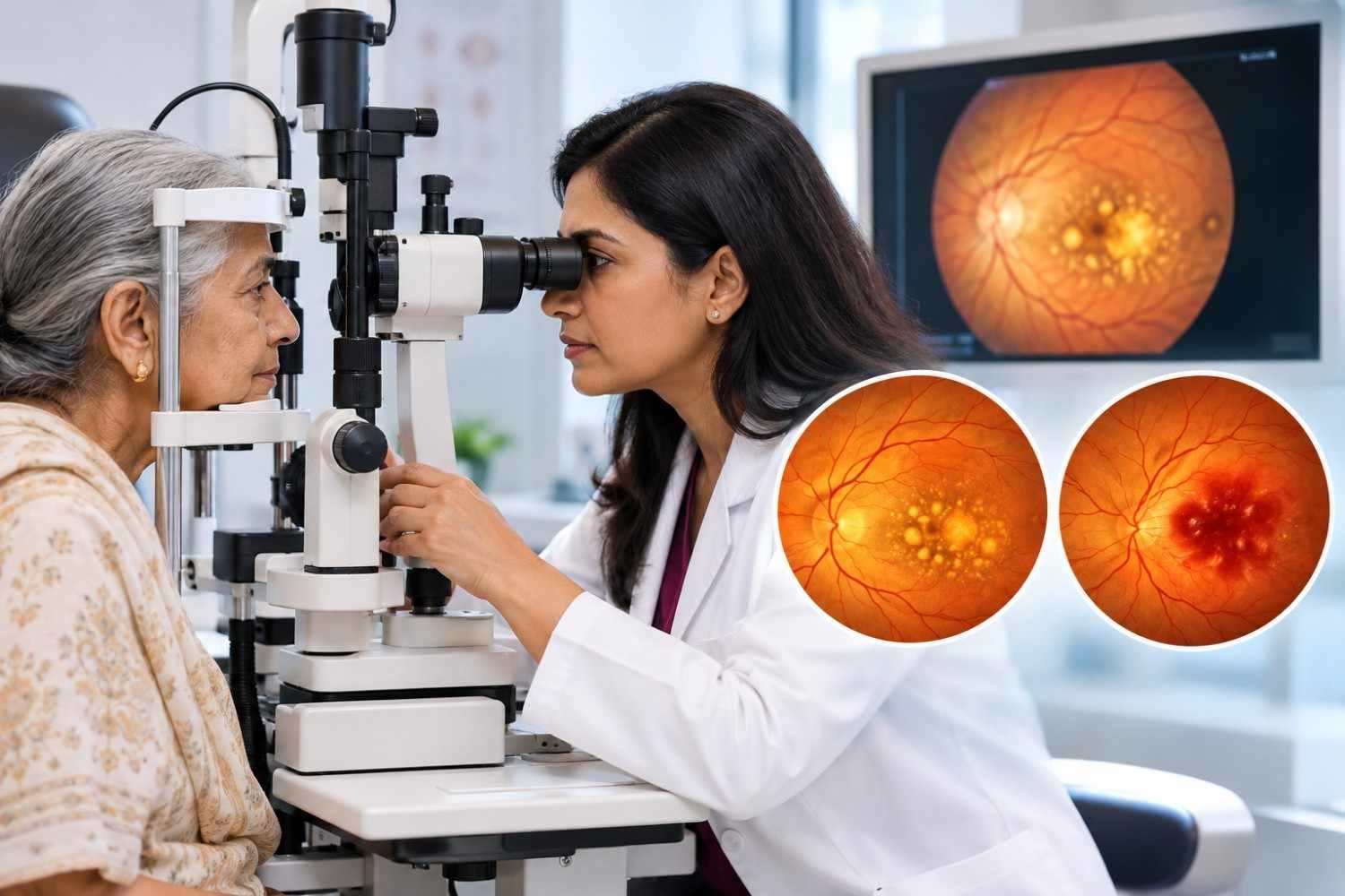
Approximately 80–85% of all AMD cases are dry AMD. It is the foundational form of the disease — all wet AMD begins as dry AMD.
What Happens in Dry AMD?
The retinal pigment epithelium (RPE) — a single layer of cells beneath the photoreceptors that supports and nourishes them — begins to malfunction with age. Metabolic waste products accumulate beneath the RPE as small deposits called drusen, visible during OCT and dilated fundus examinations.
Over time, drusen enlarge and multiply, causing the RPE cells above them to deteriorate. When RPE cells die, the photoreceptors (cone and rod cells) responsible for central vision also die. This progressive photoreceptor loss is called geographic atrophy (GA) in its advanced form, leaving corresponding blank patches in central vision.
The Four Stages of Dry AMD
| Stage | What Is Happening | Symptoms | Action |
|---|---|---|---|
| Early Dry AMD | Small drusen (< 63 microns) present under the macula. | None — completely asymptomatic. | Annual OCT monitoring; AREDS2 assessment. |
| Intermediate Dry AMD | Medium-large drusen; some RPE abnormality. | Mild central blurring; difficulty in low light; colour changes possible. | 6-monthly OCT; AREDS2 supplements if indicated; Amsler grid at home. |
| Advanced Dry AMD (Geographic Atrophy) | Large areas of RPE and photoreceptor death at the macula. | Significant central blank patch; reading and face recognition very difficult. | Close monitoring; low vision support; ongoing research treatments. |
| Advanced Wet AMD (Conversion) | Dry AMD progresses to wet AMD — abnormal blood vessels grow. | Sudden worsening — wavy lines, rapid vision loss. | URGENT Same-week anti-VEGF injection required. |
Is Dry AMD Treatable?
While there is currently no approved treatment to reverse existing dry AMD damage or halt geographic atrophy completely in India, proactive steps can help slow progression:
- AREDS2 Nutritional Supplements: Vitamins C & E, zinc, copper, lutein, and zeaxanthin reduce progression risk from intermediate to advanced AMD by ~25% in eligible patients.
- Lifestyle Modification: Stopping smoking, managing blood pressure, and adopting an antioxidant-rich diet slow progression.
- Regular OCT Monitoring: Enables early detection of conversion to wet AMD, which is highly treatable if caught early.
- Emerging Therapies: Treatments for geographic atrophy are in advanced international clinical trials and emerging approvals.
Wet AMD — The Urgent Form
Despite affecting only 10–15% of AMD patients, wet AMD is responsible for the majority of severe central vision loss from AMD — because it progresses rapidly and destructively if not treated promptly.
What Happens in Wet AMD
In wet AMD, abnormal blood vessels grow beneath the retina from the choroidal layer below — a process called choroidal neovascularisation (CNV). These new vessels are structurally fragile, leaking fluid and blood into and under the retina. This leakage distorts photoreceptors, causes macular swelling, and, if untreated, leads to scarring that permanently destroys central vision.
The progression from first leakage to significant, irreversible vision loss can occur in weeks. This is why wet AMD is treated as urgent — not as a condition that can wait for the next available appointment.
How Wet AMD Presents
Sudden Distortion
- Straight lines appear wavy, bent, or broken (metamorphopsia)
New Central Blur
- A fresh central blurry spot that was not present or was much milder before
Faded Colours
- Colours appear less vivid, duller, or altered, particularly in central vision
Size Perception Shift
- Objects in the centre of vision appear smaller or larger than they actually are
Central Dark Patch
- A new grey, dark, or blurry shadow blocking the centre of sight
Rapid Progression
- Symptoms that noticeably worsen over days to weeks rather than months
🚨 Sudden Distortion or Vision Change With Known AMD — Same-Week Evaluation
If you have known AMD — dry or wet — and notice sudden new distortion, a new central patch, or rapidly changing vision, call Raahi Netradham immediately on +91 7078082666. This may indicate conversion from dry to wet AMD or a new CNV lesion requiring urgent anti-VEGF treatment. Do not wait for your next scheduled appointment.
Treatment of Wet AMD — Anti-VEGF Injections
Anti-VEGF (anti-vascular endothelial growth factor) intravitreal injections are the gold-standard treatment for wet AMD worldwide — and have transformed the prognosis of this condition since their introduction. VEGF is the growth factor that drives abnormal vessel formation. Blocking it stops new vessel growth and reduces leakage.
Available Anti-VEGF Medications
| Anti-VEGF Medication | Generic Name | Available in India | Notes |
|---|---|---|---|
| Avastin | Bevacizumab | Yes — Widely Available | Off-label use; lower cost; widely used |
| Accentrix / Lucentis | Ranibizumab | Yes | Licensed for AMD; FDA-approved indication |
| Eylea | Aflibercept | Yes | Licensed for AMD; longer durability in many patients |
| Vabysmo | Faricimab | Limited Availability | Dual-action; newer agent; available at select centres |
Treatment Protocol Steps
Step 1: Loading Phase
- Typically 3 monthly injections to establish control of CNV leakage
Step 2: Maintenance Phase
- OCT-guided treat-and-extend or PRN (as-needed) protocol based on fluid activity
Step 3: Monitoring
- OCT at every injection visit to assess whether fluid is resolving
Step 4: Duration
- Ongoing injections over years; frequency reduces as disease stabilises
The majority of wet AMD patients treated with anti-VEGF injections achieve stabilisation of vision loss. Approximately 30–40% experience meaningful vision improvement. The key determinant of outcome — as with almost all retinal conditions — is how quickly treatment is started after the onset of leakage.
Anti-VEGF Injections for AMD at Raahi Netradham
All anti-VEGF medications for AMD — Avastin, Accentrix, and Eylea — are available in-house at Raahi Netradham on Haridwar Bypass Road, Dehradun. Dr. Mohit Garg (FVRS) and Dr. Chintan Desai (FVRSSN) perform intravitreal injections at every clinic visit, with OCT monitoring at the same session. Patients from across Uttarakhand — including Haridwar, Rishikesh, Mussoorie, and Tehri — do not need to travel to Delhi for AMD injection treatment.
Dry AMD vs Wet AMD — The Critical Difference
| Feature | Dry AMD | Wet AMD |
|---|---|---|
| Prevalence | 80–85% of all AMD cases | 10–15% of all AMD cases |
| Mechanism | Drusen accumulation → RPE atrophy → photoreceptor death | Choroidal neovascularisation → fluid/blood leakage → scarring |
| Speed of Progression | Slow — months to years | Rapid — weeks to months if untreated |
| Symptoms (Early) | None — completely silent | None initially; then sudden distortion |
| Symptoms (Advanced) | Gradual central blur; blank patch develops over years | Rapid central distortion; vision loss in weeks |
| Treatment | No curative treatment; AREDS2 may slow progression | Anti-VEGF injections — highly effective when started promptly |
| Reversible? | No — lost cells do not regenerate | Partially — leakage can stop; some vision recovery possible |
| Urgency | Monitor regularly — not an emergency unless converting | URGENT — same-week treatment when symptomatic |
| Risk of Blindness | Advanced GA can cause severe central vision loss over years | Can cause severe loss in weeks without treatment |
| Monitoring | OCT every 6–12 months depending on stage | OCT at every injection visit; monthly initially |
| Fellow Eye Risk | Fellow eye has 15–20% risk of developing wet AMD | Very high risk — fellow eye monitored closely |
Symptoms of AMD — What to Watch For
One of the most dangerous aspects of AMD is that its early stages produce no symptoms at all. Patients with early to intermediate dry AMD see normally, read normally, and drive normally. The disease is invisible to the patient until it has progressed significantly.
Symptoms When They Appear
- Central Blurring: Difficulty seeing fine detail in the centre of vision while peripheral vision remains intact
- Difficulty Reading in Lower Light: Needing brighter light or higher contrast to read comfortably
- Distorted Lines (Metamorphopsia): Straight lines appearing wavy, bent, or broken — door frames, tiles, rulers, or text lines — particularly noticeable on an Amsler grid
- Faded Colour Perception: Colours appearing less vivid, dull, or washed out in the central visual field
- Object Size Alteration: Objects in the centre of vision appearing smaller or unnaturally distorted
- Central Scotoma: A blank, dark, or grey patch blocking the absolute centre of vision
- Facial Recognition Difficulty: Central facial features appearing blurred or obscured when looking directly at someone
The Amsler Grid — A Simple At-Home Warning Tool
The Amsler grid is a simple square grid with a central dot used at home to monitor for AMD progression between clinic visits. Cover one eye, focus on the central dot from normal reading distance (with your reading glasses on), and note whether any lines appear wavy, bent, or if any section of the grid is blurry or missing.
For patients with known dry AMD, checking the Amsler grid weekly (monocularly — one eye at a time) is the most critical early-warning step. Any new distortion is the signal to call Raahi Netradham immediately rather than wait for your next scheduled checkup.
Treatment for Dry AMD — What Works, What Does Not, and What Is Coming
While there is currently no cure to reverse damage caused by dry Age-Related Macular Degeneration (AMD), proven clinical interventions can significantly slow its progression and preserve vision. Here is a clear breakdown of evidence-based options available for dry AMD.
| Intervention | Evidence Level | Who Benefits | Indian Availability |
|---|---|---|---|
| AREDS2 Nutritional Supplements | Strong Reduces intermediate-to-advanced progression risk by ~25% |
Intermediate AMD patients with specific drusen characteristics | ✅ Available Formulations sold in India; consult specialist before starting |
| Smoking Cessation | Strong Directly reduces risk and significantly slows rate of progression |
All AMD patients who smoke | ✅ Available |
| Dietary Improvement | Moderate High intake of leafy greens, fish, and coloured vegetables lowers risk |
All AMD patients | ✅ Available |
| Cardiovascular & BP Control | Moderate Systemic vascular health directly supports choroidal circulation |
All AMD patients with hypertension or cardiovascular disease | ✅ Available |
| Complement Inhibitors for Geographic Atrophy (Pegcetacoplan, Avacincaptad Pegol) |
Emerging FDA-approved in USA (2023) to slow GA growth |
Advanced dry AMD with Geographic Atrophy (GA) | ⚠️️ Limited Not yet widely available in India |
| Anti-VEGF Injections | Not Indicated No proven benefit for drusen or dry geographic atrophy alone |
Not indicated for dry AMD alone (used for Wet AMD) | N/A |
Note on Supplements: Not all over-the-counter vision vitamins contain the precise AREDS2 formula (Lutein, Zeaxanthin, Vitamin C, Vitamin E, Zinc, and Copper). At Raahi Netradham, our retina specialists evaluate your specific OCT scans to determine if you are at the exact stage of AMD where AREDS2 supplementation provides a proven benefit.
How AMD Is Diagnosed at Raahi Netradham
Accurate diagnosis and ongoing monitoring are critical for managing Age-Related Macular Degeneration (AMD). At Raahi Netradham, we utilize advanced multimodal retinal imaging to detect early structural changes, track disease progression, and guide timely interventions.
1. Optical Coherence Tomography (OCT) — The Essential Investigation
Optical Coherence Tomography (OCT) is the single most important diagnostic and monitoring tool for AMD. It provides a precise, high-resolution cross-sectional image of every layer of the macula — revealing drusen beneath the RPE, cellular deterioration, geographic atrophy, and subretinal or intraretinal fluid from choroidal neovascularisation in wet AMD.
- Performed at every retinal visit for AMD patients
- Scans compared over time to track disease progression
- Directly measures treatment response following anti-VEGF therapy
2. Colour Fundus Photography
Colour fundus photographs document the high-definition surface appearance of the macula, creating a permanent visual baseline of your retina.
- Maps drusen distribution and size changes
- Documents precise areas of RPE pigmentary alterations
- Tracks geographic atrophy expansion over months and years
3. Fluorescein Angiography (FFA)
When wet AMD is suspected or confirmed, fluorescein angiography maps the abnormal blood vessel network in the choroid using a safe diagnostic dye.
- Identifies exact location, size, and activity of CNV lesions
- Detects subtle or active vascular leakage
- Guides treatment decisions for complex or atypical CNV patterns
4. OCT Angiography (OCTA)
OCT angiography is a cutting-edge, non-invasive imaging technique that visualises retinal and choroidal blood microcirculation without requiring dye injections.
- 100% dye-free and non-invasive vascular mapping
- Detects early or subtle CNV lesions missed on standard OCT
- Monitors neovascular regression across treatment cycles
At Raahi Netradham, our eye hospital is equipped with state-of-the-art retina imaging systems to ensure swift, accurate, and painless diagnostic assessments. Led by experienced vitreoretinal specialists, we ensure that every AMD patient receives personalized, evidence-based care to protect their central vision.
How AMD Is Diagnosed at Raahi Netradham
Accurate diagnosis and ongoing monitoring are critical for managing Age-Related Macular Degeneration (AMD). At Raahi Netradham, we utilize advanced multimodal retinal imaging to detect early structural changes, track disease progression, and guide timely interventions.
1. Optical Coherence Tomography (OCT) — The Essential Investigation
Optical Coherence Tomography (OCT) is the single most important diagnostic and monitoring tool for AMD. It provides a precise, high-resolution cross-sectional image of every layer of the macula — revealing drusen beneath the RPE, cellular deterioration, geographic atrophy, and subretinal or intraretinal fluid from choroidal neovascularisation in wet AMD.
- Performed at every retinal visit for AMD patients
- Scans compared over time to track disease progression
- Directly measures treatment response following anti-VEGF therapy
2. Colour Fundus Photography
Colour fundus photographs document the high-definition surface appearance of the macula, creating a permanent visual baseline of your retina.
- Maps drusen distribution and size changes
- Documents precise areas of RPE pigmentary alterations
- Tracks geographic atrophy expansion over months and years
3. Fluorescein Angiography (FFA)
When wet AMD is suspected or confirmed, fluorescein angiography maps the abnormal blood vessel network in the choroid using a safe diagnostic dye.
- Identifies exact location, size, and activity of CNV lesions
- Detects subtle or active vascular leakage
- Guides treatment decisions for complex or atypical CNV patterns
4. OCT Angiography (OCTA)
OCT angiography is a cutting-edge, non-invasive imaging technique that visualises retinal and choroidal blood microcirculation without requiring dye injections.
- 100% dye-free and non-invasive vascular mapping
- Detects early or subtle CNV lesions missed on standard OCT
- Monitors neovascular regression across treatment cycles
At Raahi Netradham, our eye hospital is equipped with state-of-the-art retina imaging systems to ensure swift, accurate, and painless diagnostic assessments. Led by experienced vitreoretinal specialists, we ensure that every AMD patient receives personalized, evidence-based care to protect their central vision.