
Schedule Your Visit
Fill out the appointment form and our team will contact you shortly.
Glaucoma Symptoms, Causes, and Treatment
Glaucoma is a group of eye conditions in which the optic nerve is gradually damaged, often in association with raised pressure inside the eye. Because this nerve carries visual information from the eye to the brain, damage to it can cause permanent vision loss. People searching for glaucoma treatment in Dehradun usually want to know three things: how to recognise the condition, what causes it, and what can be done about it. This guide covers all three.
The most important point is that open-angle glaucoma, the most common form, usually causes no pain and no obvious change in vision in its early stages. By the time symptoms are noticed, a significant amount of optic nerve damage may already have occurred, and that damage cannot generally be reversed. This is why regular eye examinations matter more than waiting for symptoms to appear.
If you are seeking glaucoma evaluation or specialized care in Uttarakhand, the ophthalmologists at Raahi Netradham in Dehradun provide advanced diagnostic assessments and customized treatment plans.
Concerned About Glaucoma? Get Expert Evaluation at Raahi Netradham
Dehradun Location | NABH Accredited | Fellowship Specialists | Non-Contact Tonometry, Perimetry & OCT Diagnostics
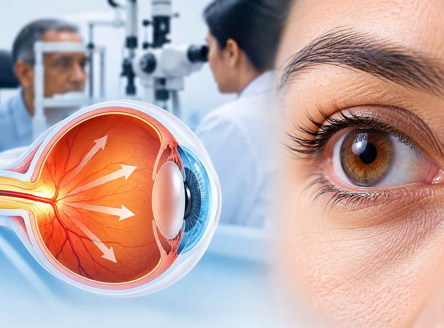
Book Your Eye Consultation in Dehradun
Get comprehensive eye care at Raahi Netradham with fellowship specialists, advanced diagnostics, and in-house treatment facilities.
What Is Glaucoma and What Causes It?
Glaucoma is a progressive optic neuropathy. The retina converts light into electrical signals, and the optic nerve carries those signals to the brain. In glaucoma, nerve fibres in the optic nerve are slowly lost, and areas of the visual field fade, usually beginning at the edges. Because this loss is gradual and the brain compensates for blind spots, central vision is often preserved until late stages.
The inside of the eye continuously produces a clear fluid called aqueous humour, which nourishes the front chamber and drains through a specialized meshwork at the drainage angle. Glaucoma develops through distinct mechanisms when this fluid dynamic or optic nerve resilience is compromised:
- • Open-angle mechanism: The drainage angle appears anatomically open, but fluid drains too slowly through the trabecular meshwork, elevating internal pressure.
- • Angle-closure mechanism: The iris physically blocks or narrows the drainage angle, either gradually over time or in an abrupt emergency blockage.
- • Secondary causes: Another medical condition, direct eye trauma, intraocular inflammation, abnormal vessel growth, or prolonged steroid medication interferes with fluid outflow.
- • Normal-tension mechanism: Optic nerve damage and visual field loss progress despite intraocular pressure remaining within statistically normal ranges.
- • Intraocular pressure (IOP) role: Higher fluid pressure stresses the sensitive optic nerve head over time; while it is the primary treatable target, pressure alone does not define the disease.
Risk factors like age, medical history, or genetics raise vulnerability, but are distinct from direct causative mechanisms. Many individuals with elevated pressure or risk factors never develop nerve damage, while others develop glaucoma with normal pressure levels.
Key Diagnostic Assessments for Glaucoma
| Diagnostic Test | What the Examination Evaluates | Clinical Importance for Diagnosis |
|---|---|---|
| Tonometry | Measures fluid pressure inside the eye (intraocular pressure / IOP). | Performed using contact instrumentation or quick non-contact air-puff methods to screen and track pressure targets. |
| Optic Nerve Examination | Detailed evaluation of the shape, cupping, and colour of the optic nerve head. | Allows direct visualization of structural cupping, thinning, or nerve tissue loss caused by glaucomatous damage. |
| Visual Field Testing (Perimetry) | Maps peripheral (side) and central field sensitivity. | Detects early, patchy blind spots and patterns of side-vision loss that patients do not consciously notice. |
| Optical Coherence Tomography (OCT) | High-resolution scan measuring retinal nerve fibre layer (RNFL) thickness. | Provides micron-level structural tracking to identify microscopic nerve loss before visual field changes appear. |
| Gonioscopy & Pachymetry | Examines the internal drainage angle and measures corneal thickness. | Determines whether angles are open or closed, and adjusts pressure readings based on corneal variations. |
Primary Risk Factors for Developing Glaucoma
Glaucoma risk increases significantly in the presence of specific genetic, anatomical, and health factors. Recognizing these helps identify when baseline screening is essential:
Age Factor (40 Years & Above)
- Risk rises steadily with increasing age, particularly after age 40
- Natural fluid outflow efficiency gradually decreases over time
- Baseline comprehensive screening is recommended for all adults by age 40
Family History & Genetics
- Significantly higher risk if a parent or sibling has glaucoma
- Genetic predisposition strongly influences optic nerve vulnerability
- Requires earlier and more frequent visual field and optic nerve reviews
Elevated Intraocular Pressure (IOP)
- The most significant and modifiable risk factor for nerve damage
- Consistent high pressure puts mechanical stress on nerve fibres
- Requires periodic tonometry to detect asymptomatic pressure spikes
Systemic Conditions & Diabetes
- Chronic conditions like diabetes affect microvascular circulation to the eye
- Increases the risk of complex forms like neovascular secondary glaucoma
- Careful systemic control complements optic nerve preservation
Long-Term Corticosteroid Usage
- Steroid eye drops, oral tablets, creams, or inhalers can induce high IOP
- Often leads to steroid-induced secondary open-angle glaucoma
- Never stop steroids unguided, but ensure regular ophthalmology monitoring
Anatomical & Refractive Variations
- Narrow angles and hyperopia (farsightedness) elevate angle-closure risk
- High myopia (nearsightedness) and thin corneas correlate with open-angle types
- Previous blunt eye trauma or surgical history can impede outflow channels
Having one or more risk factors does not mean glaucoma is inevitable; rather, it highlights the importance of regular comprehensive examinations to detect changes long before vision loss begins.
Clinical Overview of Glaucoma Types
Glaucoma is categorized based on anatomical configuration, speed of onset, and underlying medical causes:
| Glaucoma Type | Key Characteristics & Clinical Presentation |
|---|---|
| Primary Open-Angle Glaucoma (POAG) | The most common form. The drainage angle remains open, but microscopic outflow channels become inefficient. Progresses slowly and painlessly without early symptoms. |
| Angle-Closure Glaucoma | Occurs when the peripheral iris physically blocks the drainage angle. It can develop gradually (chronic) or abruptly (acute angle-closure), which is a sight-threatening emergency. |
| Normal-Tension Glaucoma (NTG) | Optic nerve cupping and visual field deficits progress despite intraocular pressure remaining within normal statistical ranges (typically ≤ 21 mmHg). |
| Congenital / Pediatric Glaucoma | Rare condition present at birth or early childhood due to abnormal drainage angle formation. Signs include watery eyes, light sensitivity, and an enlarged, cloudy cornea. |
| Secondary Glaucoma | Arises secondary to identifiable ocular or systemic issues, including eye trauma, chronic uveitis, advanced diabetes, mature cataracts, or prolonged corticosteroid use. |
Chronic Open-Angle vs. Acute Angle-Closure Glaucoma
Comparing the characteristics, onset speed, and urgent interventions required for the two major forms of glaucoma:
| Feature | Chronic Open-Angle Glaucoma | Acute Angle-Closure Glaucoma |
|---|---|---|
| Onset & Speed | Slow, silent, and progressive over years | Sudden, rapid spike in pressure within hours |
| Early Symptoms | None; no pain, redness, or early blurriness | Severe ocular pain, frontal headache, nausea, colored halos |
| Drainage Angle | Anatomically open, but functionally sluggish | Physically blocked or closed off by the iris |
| Medical Urgency | Managed with planned routine appointments | Medical Emergency: Immediate intervention required |
What Are the Treatment Options for Glaucoma?
Treatment aims to lower intraocular pressure to a safe target level to halt further optic nerve damage. While lost vision cannot generally be restored, appropriate therapy safeguards existing sight.
Prescription Eye Drops
The first-line defense for most patients. Drops either reduce aqueous humor production or enhance outflow. They must be taken consistently every single day as prescribed.
Laser Treatments
Procedures such as selective laser trabeculoplasty (SLT) improve drainage in open angles, while laser peripheral iridotomy (LPI) creates an opening in narrow or closed angles.
Glaucoma Filtration Surgery
Surgical interventions such as trabeculectomy or microscopic shunt implants create an alternate drainage pathway when medication and lasers are insufficient.
Lifelong Monitoring & Care
Glaucoma management requires ongoing evaluations. Regular visual field checks, OCT imaging, and tonometry ensure treatments are modified as soon as any change occurs.
Can Glaucoma Be Prevented? Practical Lifestyle Facts
Glaucoma cannot always be prevented because primary causes and genetics cannot be altered. However, early detection and conscious ocular protection prevent irreversible visual disability.
Early Detection as Prevention
The true foundation of glaucoma prevention is regular screening, enabling specialists to initiate pressure-lowering care before nerve fibres deteriorate.
Protective Eyewear
Wear certified eye protection during sports, construction, or high-risk manual tasks to prevent traumatic injury and secondary angle recession glaucoma.
Evidence-Based Lifestyle
No diet, herbal supplement, or eye exercise cures glaucoma. Maintain good systemic health and never replace prescribed eye drops with alternative remedies.
When Should You Consult a Glaucoma Specialist?
Understanding the boundary between routine periodic assessments and acute emergency situations ensures your vision remains protected.
Routine Clinical Assessments
Schedule a full evaluation if you are over 40, have a family history, manage diabetes, use steroid medications, or have high refractive errors.
Immediate Emergency Situations
Sudden severe eye ache, violent headache, nausea, corneal clouding, or rainbow halos around lights demand immediate emergency ophthalmology intervention.
Warning Signs of Glaucoma & Acute Attacks
When to Seek Immediate Emergency Eye Care
Contact an ophthalmologist immediately or go to the nearest emergency eye service if you notice:
Frequently Asked Questions About Glaucoma
Glaucoma Treatment in Dehradun at Raahi Netradham
Raahi Netradham is an eye hospital in Dehradun, Uttarakhand, providing comprehensive glaucoma care within its clinical specialties. The hospital features non-contact tonometry, optical coherence tomography (OCT), and perimetry among its in-house diagnostic facilities, ensuring precise detection and continuous monitoring of optic nerve health.
If you have a family history, are over 40, or have noticed changes in your side vision, scheduling an eye examination is an essential step. The ophthalmology team can evaluate intraocular pressure, assess the optic nerve and visual fields, and formulate a personalized treatment or monitoring strategy tailored to your eyes.
To arrange an examination, you can book an appointment online or contact the hospital directly at +91 7078082666. The hospital is open Monday to Saturday, 9 AM to 6 PM. If you experience sudden, severe ocular pain or abrupt vision loss, seek emergency ophthalmic care immediately.
Medical disclaimer: This article is for general educational purposes only and does not replace professional medical advice, diagnosis, or treatment. Glaucoma requires individualized assessment by a qualified ophthalmologist. Do not start, modify, or discontinue any eye medications without consulting your specialist.