
Schedule Your Visit
Fill out the appointment form and our team will contact you shortly.
How Often Should People with Diabetes Get Their Eyes Checked?
Here's a fact that many people with diabetes are never told: diabetic retinopathy can silently damage your eyesight for years before you notice any change in your vision.
Diabetic retinopathy is one of the most common complications of diabetes and one of the leading causes of preventable blindness. High blood sugar gradually damages the tiny blood vessels inside the retina, causing them to leak, swell, or close off completely. Because these changes usually occur without pain or obvious symptoms, many patients believe their eyes are healthy until the disease has already reached an advanced stage.
By the time you notice blurred vision, floaters, difficulty reading, or dark patches in your vision, diabetic retinopathy may already require treatments such as retinal laser therapy or intravitreal injections. Detecting the disease early often means it can be monitored and managed before vision is permanently affected.
The difference between finding diabetic retinopathy at an early stage and discovering it after symptoms appear can be significant. Early-stage disease may only require regular monitoring, blood sugar control, and annual retina examinations, while advanced stages often need specialised retina treatment to preserve vision.
In this guide, you'll learn exactly how often people with diabetes should get their eyes checked, what the different stages of diabetic retinopathy mean, why symptoms are not a reliable warning sign, and what happens during a comprehensive diabetic eye examination at Raahi Netradham, Dehradun.
Whether you've recently been diagnosed with diabetes or have been living with it for many years, understanding the correct eye checkup schedule is one of the most important steps you can take to protect your eyesight.
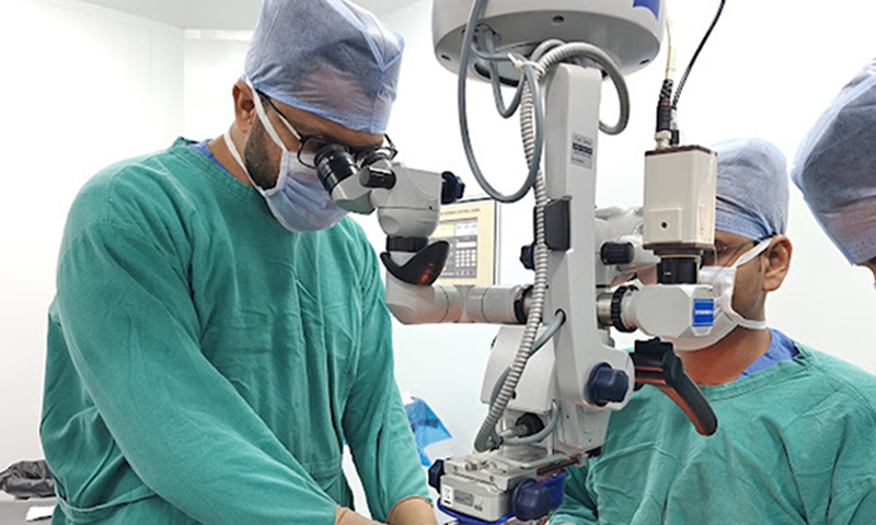
Are You Due for a Diabetic Eye Checkup?
Book your comprehensive dilated retina examination at Raahi Netradham. Our retina specialists provide advanced diabetic eye screening, OCT scans, and personalised care to detect diabetic retinopathy before it threatens your vision.
What We'll Cover
- • Why diabetes affects the eyes — explained simply
- • The exact eye checkup schedule for every diabetic patient
- • Understanding the stages of diabetic retinopathy
- • Why symptoms are not a reliable warning sign
- • What happens during a diabetic eye examination
- • Risk factors that increase the need for frequent checkups
- • Available treatment options if diabetic retinopathy is detected
- • Frequently asked questions answered by retina specialists
Why Does Diabetes Affect the Eyes?
Your retina is the light-sensitive layer at the back of your eye that allows you to see clearly. It contains thousands of tiny blood vessels that are extremely sensitive to changes in blood sugar levels.
When blood sugar remains high over months or years, these delicate blood vessels begin to leak, swell, become blocked, or grow abnormally. This condition is known as diabetic retinopathy. In its early stages it causes no pain, no vision loss, and no obvious symptoms, which is why many people don't realise anything is wrong.
As diabetic retinopathy progresses, it can lead to bleeding inside the eye, scar tissue formation, swelling of the macula (diabetic macular oedema), or even retinal detachment. At this stage, patients may experience blurred vision, floaters, difficulty reading, or permanent vision loss if treatment is delayed.
The most important thing to remember is that the damage usually starts long before you notice any change in your eyesight. Regular diabetic eye screening exists to detect these changes early—when treatment is simpler, more effective, and most likely to preserve vision.
The India Context
India has one of the world's largest diabetic populations, and diabetic retinopathy is one of the leading causes of preventable blindness among working-age adults.
Many diabetic patients in Uttarakhand have never undergone a dilated retina examination. Regular screening at Raahi Netradham helps detect diabetic eye disease before it affects vision.
Remember
Good vision does not always mean healthy eyes. Diabetic retinopathy can progress silently for years.
The Exact Checkup Schedule for Every Type of Diabetic Patient
The recommended frequency of diabetic eye examinations depends on your type of diabetes, blood sugar control, and whether diabetic retinopathy has already been detected.
| Your Situation | When to Start | How Often |
|---|---|---|
| Type 2 diabetes – newly diagnosed | At diagnosis | Every 12 months if no retinopathy is present |
| Type 1 diabetes – newly diagnosed | Within 5 years of diagnosis | Every 12 months after screening begins |
| Diabetes with no diabetic retinopathy | Ongoing | Annual retina examination |
| Mild diabetic retinopathy (NPDR Stage 1–2) | Already diagnosed | Every 6–12 months |
| Moderate to severe NPDR (Stage 3) | Already diagnosed | Every 3–4 months |
| Proliferative diabetic retinopathy (PDR) | During treatment | Every 1–3 months |
| Diabetic macular oedema | During treatment | Every 1–3 months during injections |
| Pregnancy with diabetes | First antenatal visit | Every trimester or as advised |
| After retinal laser or eye injections | Immediately after treatment | As directed by your retina specialist |
If you have diabetes and have not had a dilated retina examination within the last 12 months, schedule one as soon as possible—even if your vision feels perfectly normal. Early screening remains the best way to prevent vision loss caused by diabetic retinopathy.
What Happens If Something Is Found — Treatment Options
Finding diabetic retinopathy during a routine eye examination is not the worst outcome. Missing the condition because regular checkups were delayed is far more serious. Modern retinal treatments can effectively slow disease progression, preserve vision and, in many cases, prevent severe sight loss when started at the right time. The treatment recommended depends on the stage of diabetic retinopathy and whether the macula has been affected.
For Early-Stage Retinopathy (Stages 1–2)
During the early stages of diabetic retinopathy, the most effective treatment begins outside the clinic. Maintaining good blood sugar control, keeping HbA1c below the recommended target, controlling blood pressure, avoiding smoking and following a healthy lifestyle can significantly slow or even stabilise retinal damage. Your retina specialist will continue monitoring your eyes and recommend further treatment only if the disease progresses.
For Moderate-Severe Retinopathy (Stage 3) & PDR (Stage 4)
When diabetic retinopathy reaches an advanced stage, laser photocoagulation (Panretinal Photocoagulation or PRP) is commonly recommended. The laser treats areas of the retina that are at risk of developing abnormal blood vessels, reducing the likelihood of severe vision loss. The procedure is performed in the clinic, usually takes around 15 to 20 minutes per session, and is available at Raahi Netradham.
For Diabetic Macular Oedema
Diabetic macular oedema is most commonly treated with anti-VEGF injections, which help reduce fluid accumulation within the macula and improve central vision. The eye is numbed before the injection, making the procedure comfortable for most patients. Each treatment usually takes only 5 to 10 minutes, and several monthly injections may be required before the treatment interval is gradually extended.
For Advanced Complications
If diabetic retinopathy causes vitreous haemorrhage or tractional retinal detachment, vitrectomy surgery may become necessary to restore and protect vision. At Raahi Netradham, Dr. Mohit Garg and Dr. Chintan Desai perform advanced vitrectomy using modern 25G and 27G micro-incision systems, allowing patients in Dehradun to receive specialised retinal surgical care without travelling to Delhi.
Most diabetic eye diseases can be managed successfully when detected early. Regular retina examinations allow treatment to begin before permanent vision loss occurs, giving patients the best opportunity to maintain healthy eyesight for years to come.
What the Stages of Diabetic Retinopathy Mean for Your Checkup Frequency
Understanding the stages helps you understand why your doctor recommends different follow-up intervals. Here is what each stage means in simple language and how often you should typically have your retina examined.
Stage 1 — Mild NPDR
Annual CheckupTiny balloon-like swellings (microaneurysms) appear in the retinal blood vessels, but vision usually remains completely normal. Treatment mainly focuses on maintaining good blood sugar control, with yearly retina examinations recommended.
Stage 2 — Moderate NPDR
Every 6–12 MonthsMore retinal blood vessels become damaged or blocked, although most patients still notice no visual symptoms. Regular monitoring and strict diabetes management are essential to slow progression.
Stage 3 — Severe NPDR
Every 3–4 MonthsA large number of blood vessels become blocked, reducing blood supply to the retina. The eye begins signalling for abnormal new blood vessels to grow. Laser treatment or eye injections may be required even if vision still feels normal.
Stage 4 — Proliferative Diabetic Retinopathy (PDR)
Monthly or More FrequentlyFragile new blood vessels develop on the retina and can bleed suddenly, causing floaters, blurred vision or severe vision loss. Active treatment is required, and frequent follow-up appointments are critical to protect eyesight.
If You Are at Stage 4
Do Not Miss Follow-UpsProliferative diabetic retinopathy is a sight-threatening emergency. Missing scheduled appointments significantly increases the risk of permanent vision loss. If you have been diagnosed with PDR, arrange your next retina review without delay.
Diabetic Macular Oedema (DMO)
Usually Monthly During TreatmentDMO can occur at any stage of diabetic retinopathy. Fluid builds up in the macula, causing blurred central vision. Early treatment with anti-VEGF injections is highly effective, and close monthly monitoring is usually recommended while treatment is ongoing.
Why Follow-Up Matters
Protect Your Vision
Diabetic retinopathy often progresses silently. Regular retina checkups allow problems to be detected early, helping preserve vision with timely treatment.
Important Reminder
"Even if your eyesight feels perfectly normal, diabetic retinopathy may still be progressing. Following your recommended checkup schedule gives the best chance of preventing permanent vision loss."
— Retina Care Team, Raahi NetradhamFactors That Increase Your Risk — and Raise the Urgency
Every person with diabetes should have regular retina examinations. However, certain health conditions and lifestyle factors significantly increase the risk of developing diabetic retinopathy or make existing disease progress more rapidly. If you have any of the following risk factors, your retina specialist may recommend more frequent screening to detect retinal changes before vision is affected.
HbA1c Above 7.5% (Poorly Controlled Blood Sugar)
Persistently high blood sugar causes faster and more severe damage to the tiny retinal blood vessels, increasing the likelihood of diabetic retinopathy progression. Patients with poor diabetes control should generally undergo retina examinations every 3 to 6 months instead of waiting for an annual checkup.
Duration of Diabetes — 10+ Years
The longer you have diabetes, the greater the chance of retinal damage, even if your eyesight remains completely normal. Patients living with diabetes for more than ten years should have at least an annual retina examination, or sooner if any new visual symptoms develop.
High Blood Pressure (Hypertension)
High blood pressure works together with diabetes to accelerate damage to the retinal blood vessels. Keeping both blood pressure and blood sugar under control, along with a minimum of yearly retina screening, helps reduce the risk of vision-threatening complications.
High Cholesterol (Dyslipidaemia)
Elevated cholesterol levels contribute to fatty deposits (hard exudates) within the retina, increasing the risk of retinal damage. Annual eye examinations and appropriate cholesterol management are recommended to help protect long-term vision.
Pregnancy with Diabetes
Pregnancy can cause diabetic retinopathy to worsen much more rapidly. Women with diabetes should undergo retinal screening from their first antenatal visit and continue eye examinations during every trimester throughout pregnancy.
Kidney Disease or Previously Diagnosed Retinopathy
Diabetic kidney disease often indicates similar damage to the retinal blood vessels, while previously diagnosed diabetic retinopathy requires closer observation for progression. Most patients should have retina examinations at least every six months or follow the personalised schedule advised by their retina specialist.
Having one or more of these risk factors does not necessarily mean you will lose vision, but it does mean your eyes require closer monitoring. Following the screening schedule recommended by your retina specialist allows problems to be detected early, when treatment is most effective and vision can often be preserved.