
Schedule Your Visit
Fill out the appointment form and our team will contact you shortly.
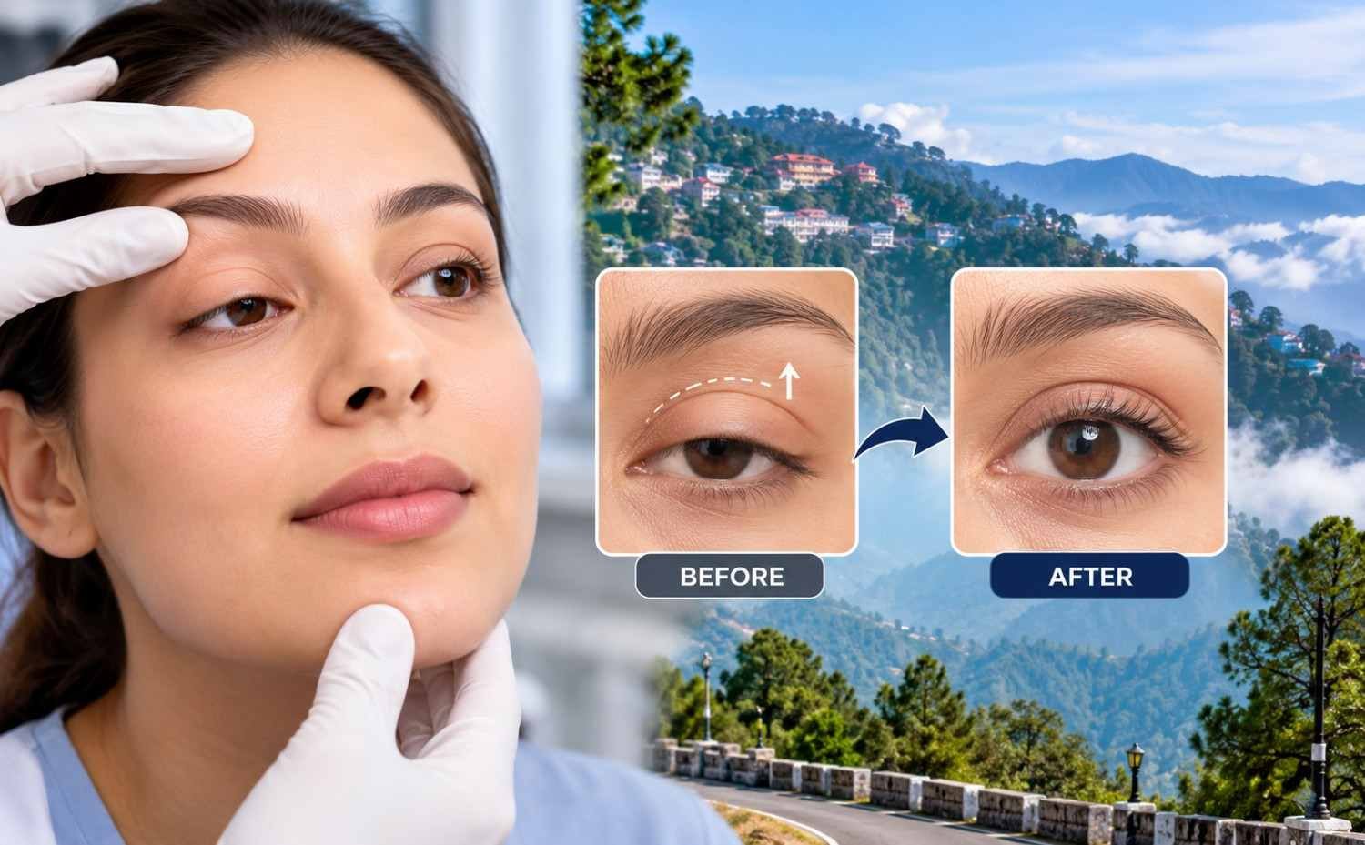
Ptosis Surgery Near Mussoorie: What to Expect Before and After Drooping Eyelid Correction
There is a particular kind of quiet frustration that comes with a drooping eyelid. Not dramatic enough to dominate a medical consultation, but persistent enough to affect every photograph, every mirror glance, and — in significant cases — every attempt to read or drive without tilting the chin upward. Patients in Mussoorie, Landour, Barlowganj, and across the surrounding hill communities have lived with this frustration for years, often assuming that the surgery they need requires Delhi.
It does not. Dr. Isha Agarwalla at Raahi Netradham on Haridwar Bypass Road in Dehradun is one of the very few FAICO-fellowship-trained oculoplastic surgeons in Uttarakhand — and the 35-kilometre road from Mussoorie to Dehradun is the distance between a drooping eyelid and a permanent correction.
This guide explains ptosis completely — what causes a drooping eyelid, what the different types of ptosis require, what surgery involves step by step, what to expect in the days and weeks after the procedure, and what patients from Mussoorie and the surrounding hill region need to know about planning the journey.
Mussoorie Patient With a Drooping Eyelid? Book a Consultation With Dr. Isha
FAICO Oculoplasty Surgeon | 60 Minutes from Mussoorie | NABH Accredited | Raahi Netradham Dehradun
Book Your Eye Consultation in Dehradun
Get comprehensive eye care at Raahi Netradham with fellowship specialists, advanced diagnostics, and in-house treatment facilities.
What Is Ptosis — And Why Doesn't It Fix Itself?
Ptosis (pronounced TOE-sis) is a drooping of the upper eyelid below its normal resting position. The upper eyelid's position is controlled primarily by the levator palpebrae superioris muscle — the 'eyelid lifter' — which originates from the back of the orbit and attaches to the upper tarsal plate and eyelid skin via a tendon-like structure called the levator aponeurosis.
When the levator muscle or its aponeurosis is weakened, stretched, or disconnected, the eyelid cannot maintain its normal height. It droops — partially or fully covering the pupil depending on severity. The patient compensates by raising the eyebrows (using the frontalis muscle as a substitute lifter), tilting the chin upward to see under the drooping lid, or developing a characteristic look of apparent fatigue or disinterest that is entirely the eyelid's doing, not the person's.
Ptosis does not resolve spontaneously. The levator muscle does not strengthen with time or exercise. Drops applied to the eye (a sympathomimetic called apraclonidine) produce only a temporary, partial elevation — useful for diagnosis, not for treatment. Surgery is the only definitive correction.
Why Mussoorie Patients Often Live With Ptosis Longer Than They Should
Ptosis in Mussoorie's hill community has historically been undertreated for two reasons: limited awareness that the condition is surgically correctable at all, and the assumption that the surgery requires a Delhi trip. Both assumptions are wrong. Ptosis is entirely correctable with a 45-60 minute procedure. And Dr. Isha Agarwalla at Raahi Netradham is 35 km from Mussoorie.
What Causes a Drooping Eyelid?
The cause of ptosis determines the surgical approach — which is why a thorough clinical assessment before any surgery recommendation is essential. Here are the main causes:
| Type of Ptosis | Cause | Who It Affects | Surgical Approach |
|---|---|---|---|
| Aponeurotic (Involutional) |
Age-related stretching or dehiscence of the levator aponeurosis — the most common type in adults | Adults over 50; often worsens gradually over years | Levator aponeurosis repair — most predictable results |
| Congenital | Underdevelopment of the levator muscle from birth; varying degrees of severity | Infants and children — present from birth or early life | Levator resection (if levator function adequate) or frontalis sling (if poor function) |
| Mechanical | Eyelid weight from tumour, inflammation, or scarring pulling the lid down | Any age — associated with an eyelid mass or structural change | Treat underlying cause; lid correction |
| Neurogenic | Nerve damage affecting levator function — 3rd nerve palsy, Horner syndrome | Any age; often sudden onset; requires neurological investigation | Neurological evaluation first; surgical correction after stability |
| Myogenic | Muscle disease — myasthenia gravis, CPEO, muscular dystrophy | Any age; often bilateral; systemic symptoms present | Medical management first; surgery in selected stable cases |
| Traumatic | Injury to the levator or its nerve supply | Any age after trauma | Assessment of residual function; 6-month wait then repair |
Important — Sudden Onset Ptosis Is a Medical Emergency
If ptosis develops suddenly — particularly if accompanied by double vision, a dilated pupil in the same eye, or headache — this may indicate a 3rd nerve palsy from an aneurysm or other neurological emergency. Any sudden-onset ptosis with these features must be evaluated urgently — call Raahi Netradham immediately or go to the nearest emergency department. Gradual ptosis developing over months or years in an otherwise healthy adult is typically age-related and not an emergency.
When Does Ptosis Need Surgery?
Not every drooping eyelid requires immediate surgery. Dr. Isha Agarwalla assesses each patient individually to determine whether and when surgical correction is appropriate.
Pupil Blockage & Functional Visual Loss
- Eyelid covers part or all of the pupil
- Obstructs the visual axis directly
- Clear indication for surgical intervention
Compensatory Posture & Chin Tilt
- Tilting chin upward continuously to see under the lid
- Causes chronic neck and shoulder strain
- Indicates significant functional ptosis
Frontalis Overaction & Brow Strain
- Continuous raising of eyebrows to lift the lid
- Causes persistent forehead creasing and brow ache
- Requires surgical elevation of the lid muscle
Pediatric Ptosis & Amblyopia Risk
- Obstructs visual axis in infants and young children
- Urgent indication to prevent amblyopia (lazy eye)
- Prevents permanent developmental vision loss
Cosmetic & Psychosocial Impact
- Noticeable drooping affecting self-confidence and presentation
- Significant impact on professional and social life
- Valid indication even when vision isn't fully blocked
When Surgery May Be Deferred
- Mild ptosis (<2mm) without functional or cosmetic strain
- Recent neurogenic or traumatic ptosis (wait 6 months)
- Myasthenia gravis requiring prior medical optimisation
At Raahi Netradham, Dr. Isha Agarwalla provides careful clinical evaluation to help patients determine the ideal timing and surgical approach for drooping eyelid correction.
What to Expect During Your Pre-Operative Assessment at Raahi Netradham
Structured Clinical Evaluation — What Happens Before Ptosis Surgery
1. External Photographs
Standardised frontal and close-up photographs in primary gaze, upgaze, and downgaze to document baseline lid position.
2. Marginal Reflex Distance (MRD1)
Measurement from corneal light reflex to upper lid margin (normal is ~4–5mm) to quantify ptosis severity accurately.
3. Levator Function Check
Measures lid excursion from downgaze to upgaze (normal >15mm) to select levator repair vs. frontalis sling surgery.
4. Eyelid Skin Crease Evaluation
Assessment of upper eyelid skin fold height and symmetry to plan natural skin crease position during surgery.
5. Bell's Phenomenon Assessment
Evaluates whether the eye rolls upward on closure to ensure corneal safety and protection post-procedure.
6. Dry Eye Screening
Evaluation of tear film quality, as corrected lid position changes surface exposure after surgery.
7. Phenylephrine Test
Sympathomimetic drop application to evaluate if Müller's muscle advancement technique is appropriate.
8. Neurological Evaluation
Thorough check to rule out underlying neurogenic or myogenic causes before planning surgical correction.
Clear Discussion & Planning
Dr. Isha discusses the surgical plan, expected symmetry, risks, and realistic outcome expectations before scheduling.
The Surgery — What Happens Step by Step
Ptosis surgery for the most common type — aponeurotic (age-related) ptosis — takes approximately 30–60 minutes under local anaesthesia. Here is how each surgical approach is performed:
1. Levator Aponeurosis Repair (Most Common — Aponeurotic Ptosis)
- Positioning & Anaesthesia: The patient is positioned comfortably on the surgical table, and local anaesthetic is injected into the upper eyelid.
- Hidden Incision: An incision is made along the natural upper eyelid skin crease — completely hiding the scar within the natural fold.
- Exposing the Muscle: The skin and orbicularis muscle are carefully opened to expose the orbital septum and levator aponeurosis.
- Muscle Reattachment: The dehisced (separated) or stretched aponeurosis is identified and reattached to the tarsal plate using absorbable sutures.
- Intra-Operative Assessment: The patient is asked to open their eyes during the procedure to assess eyelid height and symmetry in its natural position. Adjustments are made immediately.
- Closure: The skin crease is reformed, and the skin is closed with fine absorbable sutures.
2. Levator Resection (Congenital Ptosis With Moderate Function)
For congenital ptosis where levator function is present but reduced, a measured amount of the levator muscle is resected (shortened) — making the muscle more effective at lifting the eyelid. The exact amount resected is calculated based on ptosis severity and measured levator function, performed through an external (skin) or internal (conjunctival) approach.
3. Frontalis Sling (Poor Levator Function)
When levator function is very poor (<4mm excursion), the levator muscle cannot meaningfully lift the lid. A frontalis sling connects the upper eyelid directly to the frontalis (brow) muscle using a strip of fascia, silicone rod, or synthetic material — allowing the patient to elevate the eyelid smoothly by raising their brow.
| Ptosis Type | Levator Function | Surgical Technique | Anaesthesia | Duration |
|---|---|---|---|---|
| Aponeurotic (Age-related) |
Good (>10mm) | Levator aponeurosis repair | Local — patient awake | 30–45 min |
| Congenital — Moderate | Moderate (4–10mm) | Levator resection | GA in children; Local in adults |
45–60 min |
| Congenital — Severe | Poor (<4mm) | Frontalis sling | GA in children; Local in adults |
60–90 min |
| Mechanical | Any | Treat cause + lid repair | Depends on cause | Variable |
| Combined Ptosis + Blepharoplasty | Good | Aponeurosis repair + excess skin removal | Local | 60–90 min |
Book Your Ptosis Consultation With Dr. Isha Agarwalla
FAICO Oculoplasty Fellowship | NABH Accredited | 60 Minutes from Mussoorie | Raahi Netradham Dehradun
Before Your Surgery — What Mussoorie Patients Need to Prepare
Essential preparation guidelines to ensure a smooth procedure and comfortable return trip up the hill road.
Bring Your Spectacles
Bring your current spectacles — vision is assessed with current correction in place.
Disclose All Medications
Particularly blood thinners (aspirin, warfarin, clopidogrel) which should be stopped under medical supervision before surgery; also steroids and anticoagulants.
Arrange Transport
You will not be able to drive after ptosis surgery; the eyelid is partially closed with a pad or swelling immediately post-operatively; arrange a companion for the return journey to Mussoorie.
Bring Sunglasses
The eye will be light-sensitive and the eyelid area swollen; large-frame sunglasses help with comfort and privacy on the return journey up the hill road.
No Contact Lenses
Contact lenses cannot be worn in the operated eye for at least 2–4 weeks after surgery.
No Eye Makeup
Cosmetics should be stopped 48 hours before surgery.
Plan Your Accommodation
Most Mussoorie patients return to Mussoorie the same day; some prefer an overnight Dehradun stay for comfort, particularly for afternoon surgery slots.
The Day 1 Follow-Up Appointment
This is scheduled for the day after surgery and can be done at Raahi Netradham before returning to Mussoorie.
After Surgery — Recovery Week by Week
Understanding what to expect after ptosis surgery is the most important preparation a Mussoorie patient can do — because the appearance in the first days, while bruised and swollen, can be alarming if it was not anticipated.
| Timeframe | What Is Happening | What Patient Experiences | Instructions |
|---|---|---|---|
| Day 0–1 | Surgery performed; local anaesthetic wearing off | Upper eyelid swollen, possibly padded; mild ache settling | Keep head elevated; cool compresses; prescribed drops and ointment |
| Days 2–4 | Bruising peaks; swelling maximum | Purple-yellow bruising around eye and lower lid; swelling may close eye slightly | Bruising is normal; cool compresses; minimal bending/lifting |
| Days 5–10 | Bruising fading; swelling reducing | Appearance improving; sutures may still be visible | Sutures dissolve or are removed at Day 7 check |
| Days 10–14 | Most bruising gone; significant improvement | Presentable in public; eyelid height broadly correct | Light normal activity; no strenuous exercise yet |
| Weeks 4–6 | Residual swelling resolving; eyelid settling | Final position approaching; minor asymmetry may still be present | Return to full activities; final assessment at 6–8 weeks |
| Weeks 6–8 | Final result established | Eyelid at its settled height; skin crease natural; scar fading | Final photographs; spectacle prescription if needed |
Ptosis Surgery Near Mussoorie
What to Expect Before and After Drooping Eyelid Correction
There is a particular kind of quiet frustration that comes with a drooping eyelid — persistent enough to affect every photograph, every mirror glance, and every attempt to read or drive without tilting the chin upward. Dr. Isha Agarwalla at Raahi Netradham in Dehradun offers permanent surgical correction just 35 km from Mussoorie.

Planning Your Visit From Mussoorie — Practical Information
Essential route, travel time, and visit schedule details for patients traveling from Mussoorie to Raahi Netradham in Dehradun.
| Planning Item | Details for Mussoorie Patients |
|---|---|
| Distance | Approximately 35 km from Mussoorie Library Chowk to Raahi Netradham |
| Journey Time | 50–65 minutes depending on hill road conditions and Dehradun traffic |
| Route | Mussoorie-Dehradun road to Rispana Pul / Haridwar Bypass Road |
| Transport | Private car or taxi from Mussoorie (taxis available from Library Chowk and Picture Palace); shared vehicle to Dehradun then auto to clinic |
| Consultation Visit (Visit 1) | 60–90 minutes at clinic — assessment, photographs, treatment plan discussion |
| Surgery Visit (Visit 2) | Arrive at clinic; surgery 30–90 min depending on type; recovery observation 30 min; discharge same day in most cases |
| Day 1 Follow-Up (Visit 3) | Next morning — 20–30 minutes; can return to Mussoorie immediately after |
| Week 1 Check (Visit 4) | Suture check + early result assessment — 20–30 minutes |
| Week 6–8 Final Review | Final result assessment and photographs |
| Contact | Call +91 7078082666 or WhatsApp — mention you are travelling from Mussoorie for appropriate slot timing |