Dr. Isha Agarwalla is the dedicated FAICO-fellowship-trained oculoplasty specialist at Raahi Netradham — one of Uttarakhand's very few NABH-accredited eye hospitals. She is one of the very few FAICO-qualified oculoplastic surgeons practising in the state, managing the complete range of eyelid, lacrimal, and orbital conditions for patients across Dehradun, Mussoorie, Haridwar, Rishikesh, Tehri, Roorkee, and the wider Garhwal-Kumaon region.

Book Appointment
Schedule Your Visit
Fill out the appointment form and our team will contact you shortly.
Top 5 Signs You Need an Oculoplasty Specialist in Dehradun
Most people know when to see a dentist for a toothache or an orthopaedic surgeon for a bad knee. But when it comes to eye problems — particularly problems involving the eyelids, tear ducts, and structures around the eye — it is far less clear when a general eye doctor is enough and when a specialist is needed.
The answer matters. An oculoplasty specialist — a surgeon trained specifically in eyelid, lacrimal, and orbital surgery — manages conditions that a general ophthalmologist may diagnose but not be equipped to treat definitively. And several of these conditions, left in the wrong hands or left untreated, have consequences that are permanent.
This guide identifies the five clearest signs that your eye concern needs an oculoplasty specialist — specifically Dr. Isha Agarwalla (MBBS, DNB, FAICO, MNAMS) at Raahi Netradham on Haridwar Bypass Road in Dehradun. For each sign, we explain what is happening, why it matters, and what a specialist can do about it.
Think You Might Need an Oculoplasty Specialist?
Book a consultation with Dr. Isha Agarwalla at Raahi Netradham — NABH accredited, FAICO fellowship, Dehradun.
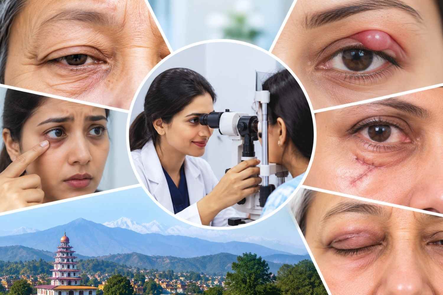
Book Your Eye Consultation in Dehradun
Get comprehensive eye care at Raahi Netradham with fellowship specialists, advanced diagnostics, and in-house treatment facilities.
First — What Is an Oculoplasty Specialist?
An oculoplasty specialist — also called an oculoplastic surgeon or ophthalmic plastic surgeon — is an ophthalmologist who has completed advanced fellowship training specifically in the surgery of the eyelids, tear drainage system, and eye socket.
In Dehradun and across Uttarakhand, Dr. Isha Agarwalla at Raahi Netradham is the dedicated FAICO-trained oculoplasty specialist. The FAICO fellowship — Fellow of the All India College of Ophthalmologists in oculoplasty — is the only oculoplasty-specific fellowship available in India, and it is what distinguishes a genuine subspecialist from a general ophthalmologist who occasionally manages eyelid concerns.
| General Ophthalmologist | Oculoplasty Specialist (Dr. Isha Agarwalla, FAICO) |
|---|---|
| Can prescribe glasses and treat common infections | Performs ptosis surgery, DCR, blepharoplasty, eyelid reconstruction |
| Can identify eyelid problems and refer | Diagnoses and treats eyelid tumours with excision and reconstruction |
| Can manage basic chalazion (eyelid cyst) | Biopsies suspicious recurrent lesions; manages eyelid cancer |
| Can manage surface eyelid lacerations | Repairs lid margin and canalicular lacerations with specialist technique |
| Can manage moderate tear duct problems with drops | Performs DCR surgery — the definitive cure for blocked tear ducts |
Sign 1 — Your Upper Eyelid Is Drooping
Sign 1: Your Upper Eyelid Is Drooping
A drooping upper eyelid — medically called ptosis — that is affecting your vision, causing you to tilt your chin upward to see under it, is worsening over time, or is present in a child is a clear sign that an oculoplasty specialist is needed.
What is happening
Ptosis occurs when the levator muscle — responsible for lifting the upper eyelid — weakens, stretches, or loses its nerve supply. The eyelid droops lower than its normal position, reducing the size of the visible eye opening.
In mild ptosis, the eyelid drops only slightly — a cosmetic concern without functional impact. In moderate to severe ptosis, the drooping eyelid partially or fully covers the pupil — blocking the visual axis and obstructing sight in the affected eye.
Why it needs a specialist
- In children — ptosis that covers the pupil during visual development causes amblyopia (lazy eye). This is a sight-threatening condition that is irreversible after age 7-8 if not treated. A child with any degree of significant ptosis should be seen urgently.
- In adults — moderate-severe ptosis causes functional visual obstruction, fatigue from constantly raising eyebrows to lift the lid, neck strain from chin-up head posture, and significant impact on appearance and confidence.
- Ptosis can be the first visible sign of a serious neurological condition — particularly sudden-onset ptosis with a dilated pupil (3rd nerve palsy — possible aneurysm) or with a small pupil (Horner syndrome — possible carotid or lung pathology). These need urgent evaluation.
What Dr. Isha Agarwalla does
Dr. Isha assesses levator muscle function, eyelid position, and visual impact, then plans the surgical technique — levator resection, Müller's muscle advancement, or frontalis sling — specific to each patient's anatomy and degree of ptosis. The result, when performed well, looks completely natural — most patients' friends cannot tell they had surgery.
💬 Dr. Isha Agarwalla — Oculoplasty Specialist, Raahi Netradham
"Ptosis surgery is one of the most rewarding procedures in oculoplasty — because the transformation is visible immediately. Patients who have been squinting, tilting, or hiding from photographs for years walk out of recovery and see a face that finally looks like them. The precision matters enormously — a millimetre in either direction changes the outcome."
💬 Real Patient Story — Dehradun
"Ek aankh ki palkh thodi neeche thi — hamesha se. Maine socha ageing hai. Phir ek behen ne notice kiya photographs mein. Dr. Isha ke paas gayi — unhone pura check kiya, bola levator muscle weak hai, surgery possible hai. Abhi operation ke baad dono aankhein ek jaisi hain. Aaj tak main yakin nahi kar paa rahi hoon ki itna simple solution tha ek cheez ke liye jo mujhe itne saalon se disturb karti thi."
— Anjali Mehta, 42, Prem Nagar, Dehradun ("One eyelid had always been slightly lower. I thought it was ageing. Then a sister noticed it in photographs. I went to Dr. Isha — she examined everything fully, said the levator muscle is weak, surgery is possible. After surgery now both eyes are the same. Even today I can't believe there was such a simple solution to something that had bothered me for so many years.")
Sign 2 — One Eye Has Been Watering Constantly for Weeks or Months
Sign 2: Persistent Watery Eye — One Side, With Discharge or Infection
If one eye has been watering constantly for more than a few weeks — particularly with discharge, crusting, or repeated infections near the inner corner — this is almost certainly a blocked tear duct, not 'dry eyes.' It needs a specialist, not more eye drops.
What is happening
Tears are produced continuously to keep the eye surface moist. They drain through tiny canals (canaliculi) at the inner corner of each eyelid into the nasolacrimal sac, then down a duct into the nose. When this drainage pathway is blocked — at any point — tears have nowhere to go and overflow down the cheek. This is epiphora — the medical term for persistent watery eye.
The most common point of blockage is the nasolacrimal duct — the final section of the drainage pathway. A blocked nasolacrimal duct causes one-sided watering, discharge (the stagnant tear pool becomes infected), and recurring swelling or infection of the tear sac (dacryocystitis).
Why eye drops are not the answer
Epiphora from a blocked tear duct is a plumbing problem — the drainage pipe is blocked. Antibiotic drops treat infection on the surface but cannot unblock the duct. Lubricating drops make no difference. The definitive treatment is a surgical procedure — DCR (dacryocystorhinostomy) — that creates a new drainage pathway bypassing the blockage.
The 5-Year Problem That Resolved in One Operation
At Raahi Netradham, Dr. Isha regularly sees patients who have had one persistently watery eye for 2, 5, even 10 years — who have used countless antibiotic drops with temporary improvement that never lasts — and who have never been referred for DCR surgery. After a single DCR procedure, the watering stops permanently. The most common thing these patients say is: "I wish someone had told me about this years ago."
What Dr. Isha Agarwalla does
After a clinical assessment confirming the blockage level, Dr. Isha performs DCR surgery — either external (small incision at the side of the nose) or endoscopic (through the nose, no external scar). A fine silicone tube keeps the new pathway open during healing and is removed after 3 to 6 months. Most patients return to normal activities within 7 to 10 days.
Sign 3 — An Eyelid Lump Has Come Back in the Same Location
Sign 3: A Recurring Eyelid Lump — Same Spot, After Treatment
A lump near the eyelid that was treated as a chalazion (blocked gland) and came back in exactly the same place — especially if it is hard, irregular, causing loss of nearby eyelashes, or is in an older adult — must be evaluated by an oculoplasty specialist. This is not just a stubborn chalazion.
What is happening — and what might be happening
Most eyelid lumps are chalazions — blocked meibomian glands that form a harmless, soft cyst. They are common and almost always benign. But there is a specific scenario that Dr. Isha Agarwalla takes extremely seriously: a lump that has been treated (incised, injected with steroid, or both) and has returned in exactly the same location.
Sebaceous gland carcinoma is a malignant tumour of the eyelid that classically mimics a recurrent chalazion. It is one of the most dangerous eyelid cancers — and one of the most commonly missed, particularly in the Indian population where it is more prevalent than in Western patients.
The red flags that demand a biopsy
- A lump in the same eyelid location that has come back after treatment — once, twice, or more
- Loss of eyelashes (madarosis) at the site of the lump — a classic warning sign of malignancy
- A lump with an irregular, indurated (hard), or ulcerated surface
- A lump that is growing rapidly or has changed in appearance
- Any new eyelid lump in a patient over 60 that has been present for more than 6 weeks
- A lump at the upper eyelid — sebaceous carcinoma is more common in the upper lid
🔴 Do Not Dismiss a Recurring Eyelid Lump
Sebaceous gland carcinoma caught early — before it spreads beyond the eyelid — is surgically curable. Detected late, it can spread to lymph nodes, orbit, and systemically. A biopsy is a minor, 10-minute procedure under local anaesthesia. It is the only reliable way to confirm or exclude malignancy. If you or a family member has a recurring eyelid lump in Dehradun — please see Dr. Isha Agarwalla at Raahi Netradham without further delay.
What Dr. Isha Agarwalla does
Dr. Isha takes a detailed history of the lump — onset, prior treatments, any change in appearance. She examines the lump and the surrounding eyelid carefully, including eyelash assessment, tarsal plate involvement, and regional lymph nodes. If there is any suspicion of malignancy, a biopsy is performed in clinic under local anaesthesia. If malignant, excision with clear margins and immediate eyelid reconstruction is planned — both in the same procedure.
Sign 4 — Excess Upper Eyelid Skin Is Blocking Part of Your Vision
Sign 4: Excess Upper Eyelid Skin Hanging Over the Pupil
When the skin of the upper eyelid becomes so heavy and loose that it droops down over the pupil and partially obstructs vision — this is not just cosmetic ageing. It is a functional visual obstruction called dermatochalasis, and blepharoplasty surgery is medically indicated.
What is happening
With age, the skin of the upper eyelid loses elasticity and excess skin accumulates, forming a heavy fold that hangs progressively lower. When this fold reaches or passes the upper pupil margin, it physically blocks the upper visual field — reducing peripheral vision, causing fatigue from eyebrow-raising to compensate, and interfering with reading, driving, and other daily activities.
This condition — dermatochalasis — is different from ptosis. In ptosis, the eyelid itself is too low. In dermatochalasis, the lid position is normal but there is so much excess skin above it that the fold overhangs the pupil. Both can co-exist — and both can be addressed surgically.
How to know if it is functional or purely cosmetic
- Does the skin of your upper eyelid visibly rest on your eyelashes?
- Do you find yourself raising your eyebrows constantly to lift the skin off your eye?
- Do you feel visual fatigue, forehead ache, or squinting by midday?
- On a visual field test, is there a reduction in the upper visual field that improves when you tape the upper lid skin upward?
If the answer to any of these is yes, your blepharoplasty is a functional procedure — removing the obstructing skin to restore the visual field. This has clinical and insurance implications distinct from cosmetic blepharoplasty performed purely for appearance.
What Dr. Isha Agarwalla does
Upper lid blepharoplasty removes the precise amount of excess skin and, where needed, underlying muscle and fat. Dr. Isha marks the incision carefully to preserve the natural eyelid crease, remove the obstruction, and maintain a natural, rested — not operated — appearance. The incision heals within the crease and becomes essentially invisible over weeks to months.
💬 Real Patient Story — Dehradun
"Parhna mushkil ho gaya tha — aankhon pe jaise bojh tha. Doctor ne bola blepharoplasty karo. Dara tha ki meri aankh kharab ho jaayegi. Dr. Isha ne sab samjhaya — kahan se skin hata jayegi, recovery mein kya hoga. 10 din mein normal ho gaya aur vision clear ho gaya. Parhna dobara achha lagne laga. Kaafi saalon baad."
— Mohan Das, 68, Rajpur Road, Dehradun ("Reading had become difficult — as if there was a weight on my eyes. The doctor said have a blepharoplasty. I was scared my eye would look wrong. Dr. Isha explained everything — where the skin would be removed, what recovery would be like. Normal in 10 days and vision cleared. Reading became enjoyable again. After many years.")
Sign 5 — You Had an Eyelid Injury Near the Lid Margin or Inner Corner
Sign 5: An Eyelid Injury — Involving the Margin or Inner Corner
If you had an eyelid cut or injury — at the lid margin (the very edge of the eyelid), near the inner corner (medial canthus), or involving significant tissue loss — and it was stitched by a general surgeon or emergency doctor, it needs an oculoplasty specialist assessment. Even if the wound 'healed', it may not have healed correctly.
Why eyelid injuries are not like other lacerations
The eyelid is not ordinary skin. It has a margin — the very edge where the upper and lower lids meet — that contains the openings of the meibomian glands, the eyelash roots, and the grey line that separates the anterior and posterior lamellae. It also has, at its inner corner, two tiny canaliculi — the drainage channels that carry tears from the eye surface into the nose.
When a laceration passes through the eyelid margin or the medial canthal area, the stakes are very different from a skin laceration elsewhere on the face:
- A lid margin wound repaired without oculoplastic technique often heals with a notch — a permanent step or gap at the eyelid edge that causes chronic lash misdirection, corneal irritation, and a visible cosmetic defect
- A canalicular laceration (cut to the tear drainage canal at the inner corner) that is not identified and repaired leads to permanent watering from that eye — epiphora that cannot be fixed with drops and requires secondary DCR surgery, which is harder and less complete than primary repair
- A wound with tissue loss requires reconstructive flap or graft surgery — not straight closure
When to seek a specialist assessment — even after initial treatment
- If you notice a notch or step at the eyelid margin after a wound has healed
- If you have persistent watering from the affected eye after an eyelid laceration — suggesting the canaliculus was cut
- If the repaired eyelid looks pulled, asymmetrical, or different from before the injury
- If lashes near the injury are growing in the wrong direction (trichiasis)
Secondary repairs — fixing inadequate primary repairs — are always more complex than getting it right the first time. But Dr. Isha Agarwalla at Raahi Netradham can evaluate even old eyelid injuries and determine whether further surgical correction would improve both function and appearance.
If the Injury Is Fresh — Come Today
An eyelid laceration involving the margin or inner corner should be evaluated by an oculoplasty specialist as soon as possible — ideally within 24-48 hours. Canalicular repair in particular must be done before the cut ends retract, making identification and repair significantly harder. Call Raahi Netradham on +91 7078082666 for same-day assessment of acute eyelid injuries.
Recognise Any of These 5 Signs? Book With Dr. Isha Agarwalla Today
NABH Accredited | FAICO Oculoplasty Fellowship | Ptosis | DCR | Blepharoplasty | Eyelid Oncology
Call +91 7078082666 | WhatsApp | raahinetradham.com
Bonus — 5 More Reasons to See Dr. Isha That People Often Miss
Beyond the top 5 signs, there are several other conditions that commonly arrive at Raahi Netradham after months or years of being managed sub-optimally elsewhere:
| Condition | Common Mistake | What Dr. Isha Does |
|---|---|---|
| Entropion — inward-turning lower eyelid rubbing lashes on cornea | Lubricating drops for 'dry eye' | Outpatient surgical correction — eyelid turned back to normal position |
| Ectropion — outward-turning eyelid causing watering and redness | Antibiotic drops for 'conjunctivitis' | Surgical correction restoring normal eyelid position and drainage |
| Thyroid eye disease — bulging eyes, lid retraction | Thyroid medication only; eye symptoms ignored | Specialist oculoplastic management — orbital decompression, lid lengthening when stable |
| Orbital fracture — sunken eye or double vision after facial trauma | Observation; not referred for surgical opinion | Assessment for surgical repair — restoring orbital volume and eye position |
| Blepharospasm — uncontrolled eyelid twitching | Told 'it will pass' or given general medication | Botulinum toxin injection — highly effective functional treatment |
Meet Dr. Isha Agarwalla — Dehradun's FAICO Oculoplasty Specialist
Dr. Isha Agarwalla | MBBS | DNB | FAICO | MNAMS
Oculoplastic Surgeon & Ocular Oncologist — Raahi Netradham Eye Clinic, Dehradun
Procedures:
ptosis correction | DCR surgery | blepharoplasty | eyelid reconstruction | eyelid tumour excision | orbital fracture repair | socket reconstruction | botulinum toxin | phacoemulsification cataract surgery with premium IOLs
Availability & Location
Available: Monday – Saturday | 9 AM – 6 PM
Address: Haridwar Bypass Road, Ajabpur Kalan, Dehradun | No referral required
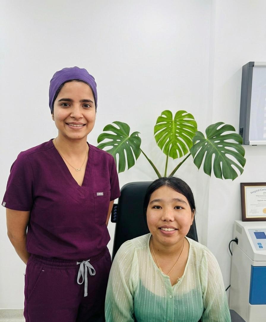
Book a Consultation With Dr. Isha Agarwalla
Oculoplasty | Cataract | Ocular Oncology — Raahi Netradham, Dehradun | NABH Accredited
Frequently Asked Questions
Ptosis cannot be improved with drops, exercises, or any non-surgical treatment. There is no medication that strengthens the levator muscle. The degree of drooping can only be reduced surgically. Whether surgery is appropriate depends on the degree of ptosis, its effect on vision, and whether it is stable — all of which Dr. Isha assesses at consultation. Mild ptosis not affecting vision may be observed. Any ptosis affecting vision, or any ptosis in a young child, warrants surgical consideration.
Yes — DCR surgery is effective regardless of how long the tear duct has been blocked. The procedure creates a new drainage pathway, bypassing the blocked section entirely, so the duration of the blockage does not affect surgical success. Patients who have had watery eyes for 10 years have the same surgical outcomes as those who have had it for 6 months. The most common thing patients say after DCR is that they wish they had done it years earlier.
You cannot reliably distinguish a chalazion from a sebaceous gland carcinoma by appearance alone — not even experienced clinicians can do so reliably without a biopsy. The key warning signs are recurrence in the same location, loss of lashes at the site, hard or ulcerated surface, and patient age over 50. If any of these are present, Dr. Isha will take a biopsy — a 10-minute procedure under local anaesthesia that provides a definitive answer. Do not accept 'it's just another chalazion' without a biopsy if the lump keeps coming back.
Yes. No referral is required. Patients from anywhere in Dehradun or Uttarakhand can book directly with Dr. Isha Agarwalla by calling +91 7078082666, WhatsApp, or walking in to Raahi Netradham on Haridwar Bypass Road, Ajabpur Kalan, Dehradun. Walk-in patients for acute eyelid injuries are seen immediately on priority.
Functional oculoplasty surgery — procedures performed to restore vision or treat a medical condition — is typically covered by health insurance and mediclaim policies in India, subject to policy terms. This includes ptosis correction that affects vision, DCR surgery for blocked tear ducts, eyelid reconstruction after injury or tumour removal, and orbital fracture repair. Purely cosmetic procedures (blepharoplasty for appearance only, aesthetic fillers) are typically not covered. Raahi Netradham's team can assist with insurance documentation and pre-authorisation.