
Schedule Your Visit
Fill out the appointment form and our team will contact you shortly.
What Tests Are Done During a Retina Examination? A Patient's Guide
Booking a retina examination for the first time — whether for yourself or for an elderly parent who has just been diagnosed with diabetic retinopathy — often comes with a quiet uncertainty: what exactly is going to happen? What are they going to do to my eyes? Is it going to hurt? How long will it take? Will I be able to drive home?
These are completely reasonable questions, and not knowing the answers can make the appointment feel more daunting than it needs to be. The truth is that most retina tests are painless, non-invasive, and take only a few minutes each.
This guide explains every test performed during a retina examination at Raahi Netradham in Dehradun — what each one involves, what the doctor is looking for, whether it is uncomfortable, and how long it takes.
By the time you finish reading, a retina appointment will feel like exactly what it is: a thorough, important, and largely comfortable experience.
Ready to Book Your Retina Examination in Dehradun?
All retina tests are available in-house at Raahi Netradham — one building, one visit.
What We'll Cover
- • Why retina examinations involve multiple tests
- • Test 1 — Visual acuity
- • Test 2 — Intraocular pressure (IOP)
- • Test 3 — Dilating drops
- • Test 4 — OCT scan (Optical Coherence Tomography)
- • Test 5 — Dilated fundus examination
- • Test 6 — Fundus photography
- • Test 7 — Perimetry (visual field testing)
- • Test 8 — Fluorescein angiography
- • Test 9 — B-scan ultrasound
- • Test 10 — Amsler grid (at-home self-test)
- • Which tests will I have at my appointment?
- • What to bring and how to prepare
- • FAQs — answered honestly
Complete Retina Testing Under One Roof
Every test described in this guide is available in-house at Raahi Netradham — including OCT, fundus photography, perimetry, fluorescein angiography, B-scan ultrasound, and slit lamp examination.
One Building. One Visit.
Patients from across Dehradun, Mussoorie, Haridwar, Rishikesh, Tehri, and the wider Garhwal region can receive their complete retina workup in one building, in one visit, without needing to go anywhere else.
Why Retina Examinations Involve Multiple Tests
The retina is a thin, light-sensitive layer no thicker than a piece of cling film, lying at the back of the eye across a surface roughly the size of a postage stamp. It contains millions of light-sensitive cells and an intricate network of blood vessels — and conditions affecting it can be microscopic in their early stages.
No single test reveals everything. Each test gives the specialist a different kind of information — like the different views on a medical scan. Together, they create a complete picture of your retinal health. A doctor relying on just one test might miss what another test would have caught clearly.
This is also why the best retina examinations happen at clinics with all the tests available in-house — so the complete picture can be assembled in a single visit, without sending patients to a different diagnostic centre between appointments.
Comprehensive Retinal Evaluation & Diagnostic Tests
An accurate diagnosis of retinal and ocular conditions relies on advanced diagnostic tools and precise evaluations. Here is a breakdown of the key eye tests conducted during a complete retinal examination:
Visual Acuity Test
What it is: Standard eye chart reading to measure clarity of vision for each eye.
Purpose: Detects changes in vision and sets a baseline for treatment outcome tracking.
Duration: 2–3 mins | PainlessIntraocular Pressure (IOP / Tonometry)
What it is: Fluid pressure measurement inside the eye via air-puff or applanation probe.
Purpose: Checks for glaucoma, monitors steroid drop effects, and ensures safety during anti-VEGF injections.
Duration: Less than 1 min | Mild surprise, no painDilating Drops (Mydriasis)
What it is: Eye drops used to widen the pupil from 2–4mm to 7–8mm.
Purpose: Allows the specialist to examine the peripheral retina for tears and diabetic changes.
Effect Duration: 3–4 hours (blurry near-vision & light sensitivity)OCT Scan (Optical Coherence Tomography)
What it is: Non-contact 3D cross-sectional imaging of retinal layers using light waves.
Purpose: Essential for detecting AMD, diabetic macular oedema, macular holes, and fluid resolution post-treatment.
Duration: 3–5 mins | Completely painlessDilated Fundus Examination
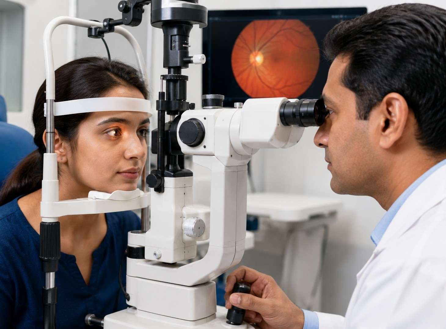
What it is: Direct clinical examination of the retina, optic disc, and vitreous using high magnification.
Purpose: Identifies retinal tears, detachments, hemorrhages, and optic nerve abnormalities.
Duration: 10–15 mins | Bright light discomfortFundus Photography
What it is: High-resolution digital imaging of the macula, optic disc, and retinal blood vessels.
Purpose: Creates a baseline to track subtle progress or disease changes over time.
Duration: 2–5 mins | Camera flash effectPerimetry (Visual Field Testing)
What it is: Automated light-response test mapping full central and peripheral vision.
Purpose: Detects side-vision loss caused by glaucoma, optic nerve disease, or brain lesions.
Duration: 15–20 mins (both eyes) | Requires focusFluorescein Angiography (FFA)
What it is: Contrast dye imaging of retinal blood flow via IV injection in the arm.
Purpose: Pinpoints blood vessel leakage, blockages, and abnormal growth in AMD or diabetic retinopathy.
Duration: 15–20 mins | Specialist diagnostic testB-Scan Ultrasound
What it is: High-frequency sound wave imaging through closed eyelids.
Purpose: Evaluates the retina when dense cataracts or vitreous hemorrhage block clear light view.
Duration: 5–10 mins | Gentle eyelid probeAmsler Grid (At-Home Self-Test)
What it is: Grid pattern test to monitor distortion in central vision.
Purpose: Serves as an early home-warning test for wet AMD and macular disorders.
Usage: Weekly home test | 1 minImportant Instructions Before Your Appointment
Due to dilating eye drops, you will experience temporary blurriness and sensitivity to light for 3–4 hours. Do not drive yourself. Please bring a pair of sunglasses and arrange a companion or alternate transport for your return home.
All Retina Tests Available In-House at Raahi Netradham, Dehradun
OCT, Fundus Photography, Perimetry, Fluorescein Angiography, and B-Scan — All under one roof in a single visit.
Call: +91 7078082666 | WhatsApp | raahinetradham.com
Which Tests Will I Have at My Retina Appointment?
Not every test is performed at every visit. The combination depends on your condition, your symptoms, and what the retina specialist is monitoring. Here is a simple guide to the tests you may typically have.
| Your Situation | Tests You Will Typically Have |
|---|---|
| First-ever retina appointment — routine check | Visual acuity, IOP, dilating drops, OCT, dilated fundus exam, fundus photography |
| Diabetic retina screening | Visual acuity, IOP, dilating drops, OCT, dilated fundus exam, fundus photography — every visit |
| Sudden floaters, flashes, possible detachment | Dilating drops, dilated fundus exam including the peripheral retina with indirect ophthalmoscopy, and B-scan if needed |
| Macular degeneration monitoring | Visual acuity, OCT, Amsler grid review, and fundus photography for comparison |
| Wet AMD or DMO — on anti-VEGF injections | Visual acuity and OCT every visit, with angiography performed periodically |
| Glaucoma or optic nerve concern | Visual acuity, IOP, OCT (RNFL), and perimetry |
| Uveitis monitoring | Visual acuity, IOP (especially if on steroid drops), slit lamp, OCT, and fundus photography |
| Dense cataract — pre-op retina evaluation | B-scan ultrasound to check the retina through the dense lens |
| Retinal vascular disease | OCT, fundus photography, and often fluorescein angiography to map retinal perfusion |
Your Tests Are Individualised
Your retina specialist will decide which tests are necessary based on your symptoms, medical history, diagnosis, and previous results. You may not need every test listed above during a single appointment.
What to Bring and How to Prepare for Your Retina Examination
What to Bring
- Your glasses or contact lenses — the visual acuity test is done with your current correction in place
- Sunglasses — your eyes will be light-sensitive after dilation. Wrap-around or large-frame sunglasses are ideal
- A companion if possible — someone who can drive you home or accompany you on public transport
- Your health insurance card and policy details if you are making a cashless or reimbursement claim
- Any previous eye reports, OCT scans, or fundus photographs from previous examinations — these allow Dr. Mohit or Dr. Chintan to compare directly
- A list of all current medications — especially blood thinners, diabetes medications, and any eye drops
What to Expect
- Allow 60 to 90 minutes for a first appointment — mostly waiting for the drops to dilate the pupils
- You will not be able to read or use your phone comfortably for 3 to 4 hours after the examination
- You will be able to see in the distance well enough to walk, but bright light will be uncomfortable
- Follow-up appointments are typically 30 to 45 minutes
Do I Need a Referral?
No. Patients from anywhere in Dehradun, Haridwar, Rishikesh, Mussoorie, Tehri, or elsewhere in Uttarakhand can book a retina examination directly at Raahi Netradham on Haridwar Bypass Road — without a letter from another doctor.
Simply call, WhatsApp, or walk in and describe your reason for visiting. Our retina team can guide you regarding the appropriate examination and diagnostic tests based on your symptoms and medical history.