
Schedule Your Visit
Fill out the appointment form and our team will contact you shortly.
When Should You Visit Dr. Chintan Desai in Dehradun? A Complete Patient Guide
Some eye conditions give you weeks to decide. Others give you hours.
Dr. Chintan Desai, Co-Founder and Senior Vitreoretina Surgeon at Raahi Netradham on Haridwar Bypass Road, treats both kinds — from the urgent retinal detachment that cannot wait past tomorrow, to the diabetic patient who needs an annual OCT scan even when their vision feels completely normal.
Understanding which category your symptoms fall into is the most important first step. This guide answers that question directly, for every condition Dr. Chintan treats.
Whether you are a patient in Dalanwala dealing with sudden floaters, a diabetic resident of Prem Nagar who has never had a retina check, or a visitor from Mussoorie whose eye test revealed a swollen optic disc — this guide will tell you what to do and how quickly to do it.
Experiencing Sudden Vision Changes Right Now?
Call Raahi Netradham immediately — Dr. Chintan Desai's team sees retinal emergencies the same day.
Expert vitreoretinal care, advanced OCT diagnostics, and emergency retinal treatment under one roof in Dehradun, Uttarakhand.
Who Is Dr. Chintan Desai?
Co-Founder & Senior Vitreoretina Surgeon providing advanced retinal care and neuro-ophthalmology at Raahi Netradham Eye Clinic, Dehradun.
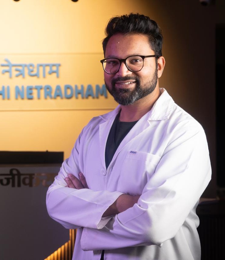
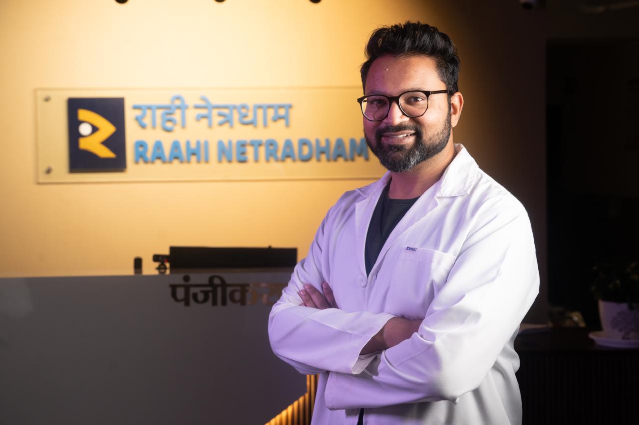
Dr. Chintan Desai
Co-Founder & Senior Vitreoretina Surgeon — Raahi Netradham Eye Clinic, Dehradun (NABH Accredited)
Dr. Chintan Desai is the Co-Founder and Senior Vitreoretina Surgeon at Raahi Netradham — one of Uttarakhand's very few NABH-accredited eye hospitals. He holds an MBBS, DNB in Ophthalmology, FICO (Fellow of the International Council of Ophthalmology), and FVRSSN (Fellowship in Vitreoretinal Surgery from Sankara Nethralaya, Chennai — one of Asia's top retina centres). He trained at the Regional Institute of Ophthalmology, Kolkata; Sankara Nethralaya, Chennai; and Sankaradeva Nethralaya, Guwahati.
His clinical expertise covers the full spectrum of vitreoretinal surgery — retinal detachment repair, macular surgery, diabetic retinopathy management, anti-VEGF injections, and complex cataract surgery with premium IOLs. He also manages neuro-ophthalmic disorders including optic neuropathy, papilloedema, visual field defects, and diplopia — conditions that sit at the intersection of eye and brain, requiring a depth of subspecialty training that is rare in Uttarakhand.
His work in macular surgery has been recognised with multiple national awards. He is an active contributor to rural eye care through the Shree Om Foundation, organising mobile eye camps and vision centres across Garhwal's most remote communities.
Detachment, Macula & Retina Care
Retinopathy & Intravitreal Injections
Optic Nerve & Vision Field Care
Sankara Nethralaya Fellowship Trained
At a Glance
Quick summary of credentials, subspecialty focus, surgical technology, and clinic details for Dr. Chintan Desai at Raahi Netradham Eye Clinic, Dehradun.
| Detail | Information |
|---|---|
| Designation | Co-Founder & Senior Vitreoretina Surgeon |
| Qualifications | MBBS, DNB (Ophthalmology), FICO, FVRSSN |
| Training | RIO Kolkata | Sankara Nethralaya Chennai | Sankaradeva Nethralaya Guwahati |
| Primary Focus | Vitreoretinal Surgery, Neuro-Ophthalmology, Complex Cataract |
| Hospital | Raahi Netradham Eye Clinic, Dehradun (NABH Accredited) |
| Surgical Tech | 25G / 27G Minimally Invasive Vitrectomy Systems |
| Serves | Dehradun, Mussoorie, Haridwar, Rishikesh, Tehri, Roorkee, Vikasnagar and all of Uttarakhand |
| Availability | Monday – Saturday, 9 AM to 6 PM |
| Booking | No referral needed — call, WhatsApp, or walk in |
Dr. Chintan Desai — Co-Founder, Raahi Netradham
"My mission at Raahi Netradham is simple: every patient who walks through our door should receive the same quality of care they would get at India's best eye hospital — without having to travel to Delhi to get it."
What Dr. Chintan Desai Treats — The Complete List
Dr. Chintan Desai provides comprehensive, specialized treatment for complex eye conditions, spanning subspecialty vitreoretinal surgery, medical retina, neuro-ophthalmology, and advanced cataract procedures.
Vitreoretinal Surgery
• Retinal detachment: Pneumatic retinopexy, scleral buckling, and pars plana vitrectomy
• Macular hole: Vitrectomy with internal limiting membrane (ILM) peeling
• Epiretinal membrane: Vitrectomy with membrane peeling
• Vitreous haemorrhage: Diagnostic and surgical management
• Tractional retinal detachment: Diabetic and non-diabetic
• Giant retinal tear: Complex vitreoretinal surgery
Retinal Vascular & Medical Conditions
• Diabetic retinopathy: All stages, screening through treatment
• Diabetic macular oedema: Anti-VEGF injections and laser
• Retinal vein occlusion: Central and branch, anti-VEGF and laser
• Retinal artery occlusion: Urgent management
• Age-related macular degeneration (AMD): Dry and wet forms
• Anti-VEGF intravitreal injections: Avastin, Accentrix, Eylea
• Retinal laser photocoagulation: Focal, grid, PRP
Neuro-Ophthalmic Disorders
• Optic neuritis: Inflammation of the optic nerve
• Ischaemic optic neuropathy: Loss of blood supply to the optic nerve
• Papilloedema: Swollen optic disc from raised intracranial pressure
• Visual field defects: Optic nerve or brain origin
• Diplopia (double vision): Cranial nerve palsies and other causes
• Horner syndrome: Drooping lid with small pupil
• Unexplained vision loss: With normal eye examination
Complex Cataract Surgery
• Phacoemulsification: With premium IOLs (toric, multifocal, trifocal, EDOF)
• Diabetic cataract surgery: In patients with co-existing retinal disease
• Dense cataracts: Brunescent and white cataracts requiring complex extraction
• Post-vitrectomy cataract surgery: Technically demanding cases
Advanced Subspecialty Care at Raahi Netradham
From emergency vitreoretinal procedures to complex neuro-ophthalmic evaluations and customized cataract surgery, every treatment plan is guided by advanced diagnostics and evidence-based protocols under the personal care of Dr. Chintan Desai.
Master Urgency Guide — When to See Dr. Chintan and How Fast
Use this table to determine how urgently you need to book. For any doubt about urgency, call Raahi Netradham and describe your symptoms — the team will guide you.
| Your Symptom or Situation | Urgency | Action |
|---|---|---|
|
Dark curtain or shadow spreading across part of vision
|
🚨 Same day — call now | Possible retinal detachment — emergency |
|
Sudden large increase in floaters — new this week
|
🚨 Same day | Possible retinal tear or early detachment |
|
Floaters + flashes of light together — new
|
🚨 Same day | High-risk vitreoretinal traction — urgent evaluation |
|
Sudden significant vision loss in one eye
|
🚨 Same day | Multiple urgent causes — vascular, retinal |
|
Red or dark haze filling your vision
|
🚨 Same day | Possible vitreous haemorrhage |
|
New double vision — appeared recently
|
⚠️ Within 48 hours | Cranial nerve palsy — needs neuro-ophthalmic evaluation |
|
Swollen optic disc found at eye examination
|
⚠️ Same/next day | Possible papilloedema — brain imaging needed urgently |
|
Straight lines look wavy or bent
|
⚠️ Within 1 week | Macular condition — AMD or epiretinal membrane |
|
Blank patch in centre of vision
|
⚠️ Within 1 week | Macular condition — OCT scan needed |
|
Diabetic — no retina check in past 12 months
|
📅 Routine — book now | Annual diabetic retina screening — even with normal vision |
|
High myopia (above -6) — no dilated retina exam
|
📅 Routine — book soon | Annual dilated exam for thin retina risk |
|
Already on anti-VEGF injections, due for next dose
|
📅 As scheduled | Follow your treatment schedule — do not delay injections |
|
Complex cataract with known retinal co-disease
|
📅 Planned | Co-ordinated surgical planning with Dr. Chintan |
|
Optic disc changes on previous OCT — monitoring
|
📅 As directed | Ongoing surveillance as per specialist advice |
Don't wait if your vision has changed suddenly. Contact Raahi Netradham and describe your symptoms so the team can guide you on the appropriate next step.
Diabetic Eye Disease — Even When You Feel Fine
Diabetic retinopathy is responsible for a significant and growing proportion of preventable blindness in Uttarakhand. And the most dangerous thing about it is how silent it is in its early stages.
Patients in Clement Town, Saharanpur Chowk, Raipur, and across Dehradun who have had diabetes for five, ten, or fifteen years and whose vision still feels perfectly sharp may already have Stage 2 or Stage 3 retinopathy developing in their retinal blood vessels — completely invisible without an OCT scan and dilated examination.
| Diabetic Patient Profile | Recommended Frequency | What Dr. Chintan Checks |
|---|---|---|
| Type 2 diabetic — newly diagnosed | At diagnosis — then annually | Baseline retinal photograph + OCT + dilated exam |
| Diabetic — no retinopathy detected | Once a year minimum | Dilated exam + OCT — looking for microaneurysms, hard exudates |
| Mild to moderate NPDR detected | Every 6 months | Monitoring progression, planning treatment timing |
| Severe NPDR — high proliferation risk | Every 3–4 months | Laser or injection treatment, close monitoring |
| PDR (proliferative) or active DMO | Monthly during treatment | Anti-VEGF injections, monitoring treatment response |
| Diabetic + poor HbA1c control | Every 3–6 months | Higher risk — more frequent surveillance |
At Raahi Netradham, every diabetic patient is assessed with OCT imaging at every visit — not just a dilated exam. OCT detects diabetic macular oedema in its earliest, most treatable stage, before it causes any visual blurring.
💬 Real Patient Story — Dehradun
Arun Rawat, 56, Prem Nagar, Dehradun:
"8 saal se diabetes hai. Doctor ne glasses ki baat boli, aankhon ki baat kabhi seriously nahi hua. Ek dost ne Raahi ka naam suggest kiya. Dr. Chintan ne OCT kiya — bola Stage 2 retinopathy hai aur ek jagah fluid bhi hai macula mein. Mujhe kuch pata nahi tha. Treatment ke baad stable hai. Ab main har 6 mahine jaata hoon. 8 saal barbaad kar diye, ab koi risk nahi lena."
("I've had diabetes for 8 years. My doctor mentioned glasses but never seriously discussed my eyes. A friend suggested Raahi. Dr. Chintan did an OCT — said Stage 2 retinopathy and some fluid in the macula. I had no idea. After treatment it's stable. Now I go every 6 months. I wasted 8 years, now I'm not taking any risk.")
Macular Conditions — Subtle Symptoms, Significant Importance
The macula is the tiny central area of the retina responsible for reading, recognising faces, and all fine-detail vision. Macular conditions affect central vision — and because they are subtle in their early stages, they are often dismissed or attributed to tiredness or needing new glasses.
Macular Degeneration (AMD)
Age-related macular degeneration is the leading cause of central vision loss in adults over 60 in high-income countries — and increasingly common in India as the population ages. Wet AMD, where abnormal blood vessels grow under the retina and leak fluid, progresses rapidly if untreated.
Key Symptoms to Watch For:
- • Straight lines that look wavy, bent, or distorted
- • A greyed-out or blank patch in the centre of your visual field
- • Difficulty reading, recognising faces, or watching television
Treatment
Anti-VEGF Injections (Avastin, Accentrix, Eylea) — highly effective when started early.
Macular Hole
A full-thickness break in the central macula — typically causing a small blank spot in the very centre of vision and distortion in close work.
Epiretinal Membrane (Macular Pucker)
A thin sheet of scar-like tissue that grows across the macula, causing it to wrinkle. The primary symptom is distortion — straight lines that appear wavy or rippled, and reduced clarity of central vision.
Neuro-Ophthalmic Disorders — When the Eye Problem Is Actually in the Brain
This is the subspecialty within Dr. Chintan's practice that most surprises patients — because it deals with vision problems that originate not in the eye itself, but in the optic nerve or the brain.
When a patient has blurry vision and their eye examination looks largely normal, the cause often lies somewhere along the visual pathway — the optic nerve, the optic chiasm, or the visual cortex in the brain. This is neuro-ophthalmology territory.
| Neuro-Ophthalmic Symptom | Possible Cause | Urgency |
|---|---|---|
| Sudden vision loss in one eye — eye looks normal | Optic neuropathy, vascular event | 🚨 Same day — emergency |
| Swollen optic disc found at routine check | Papilloedema — raised brain pressure | ⚠️ Same/next day — imaging needed |
| New double vision — one eye or both | Cranial nerve palsy — 3rd, 4th or 6th nerve | ⚠️ Within 24-48 hours |
| Double vision + drooping lid + large pupil | 3rd nerve palsy — possible aneurysm | 🚨 Emergency — go immediately |
| Missing patches in visual field — consistent pattern | Optic nerve or brain lesion | ⚠️ Within 1 week |
| Drooping lid + small pupil (Horner syndrome) | Carotid dissection or lung tumour | ⚠️ Within 1 week — imaging needed |
| Vision loss with jaw pain or scalp tenderness | Giant cell arteritis — GCA | 🚨 Emergency — steroids within hours prevent blindness |
| Eyes that don't track together | Brainstem, MS, or cranial nerve cause | ⚠️ Within 1 week |
Comprehensive In-House Evaluation
At Raahi Netradham, Dr. Chintan Desai assesses neuro-ophthalmic conditions with in-house OCT of the optic nerve fibre layer, visual field testing (perimetry), fundus photography, and coordinates neuroimaging with radiology where needed. Patients from Dehradun and across the Garhwal region no longer need to travel to Delhi for this level of neuro-ophthalmic evaluation.
Complex Cataract Surgery With Co-Existing Retinal Disease
Not all cataract surgery is straightforward. Patients who have both a significant cataract and a retinal condition — particularly diabetic patients, those with age-related macular degeneration, or patients who have previously undergone retinal surgery — need their cataract managed by a surgeon who understands the retinal implications.
Cataract in a Diabetic Patient
The retina must be evaluated before surgery, diabetic macular oedema pre-treated if needed, and the surgical plan adjusted for the co-existing retinal condition.
Cataract After Vitrectomy
The eye is anatomically altered — lens power calculation is more challenging, and the surgery itself requires a different technique.
Dense Cataract Hiding Retinal Pathology
When a cataract is too dense to see through, an ultrasound B-scan is used to thoroughly evaluate the retina prior to surgery.
IOL Selection With Macular Disease
Standard IOL power formulas may need modification for eyes with underlying macular pathology affecting vision.
Dr. Chintan Desai performs phacoemulsification with premium IOLs — including toric, multifocal, trifocal, and EDOF lenses — for complex cases where both cataract and retinal expertise are needed in the same surgeon's hands.
What to Expect at Your First Appointment With Dr. Chintan Desai
Whether you are coming for an urgent evaluation or a planned first consultation, here is exactly what a visit with Dr. Chintan looks like at Raahi Netradham.
Registration & History
Detailed history of onset, nature, and symptom progression; medical history (diabetes, BP, neurological conditions); and current medications (blood thinners, eye drops).
Visual Acuity
A standard check to evaluate how clearly you see with each eye individually.
Intraocular Pressure
A quick, painless check crucial for retinal vascular conditions and neuro-ophthalmic evaluations.
Dilating Drops
Pupils are widened to allow a full retinal examination. Takes 15–20 mins.
OCT Scan
High-resolution, in-house imaging of the retina and optic nerve. Painless, takes only 5 minutes, and provides details invisible on direct examination alone.
Dilated Fundus Exam
A comprehensive examination of the retina, macula, optic disc, and vitreous body using a specialist lens and light source.
Visual Field Testing
Perimetry testing conducted where relevant for neuro-ophthalmic or retinal condition assessments.
Explanation of Findings
Dr. Chintan explains the diagnostic results clearly, ensured in easy-to-understand terms you can take home.
Personalised Treatment Plan
Medical, laser, injection, or surgical options are discussed openly, including expected visual outcomes and recovery timeline.